. 2016 Mar 11;11(3):e0150411.
doi: 10.1371/journal.pone.0150411. eCollection 2016.

A Gain-Of-Function Mutation in the Plcg2 Gene Protects Mice from Helicobacter felis-Induced Gastric MALT Lymphoma

Affiliations

A Gain-Of-Function Mutation in the Plcg2 Gene Protects Mice from Helicobacter felis-Induced Gastric MALT Lymphoma

Jennifer Gossmann et al. PLoS One. .

Abstract

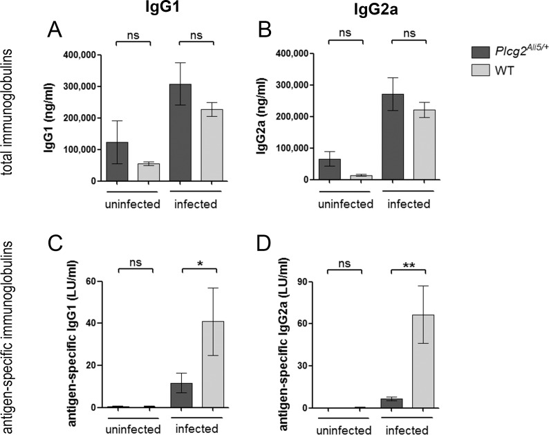
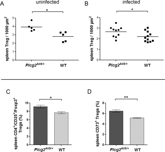
Gastric mucosa-associated lymphoid tissue (MALT) lymphomas develop from a chronic Helicobacter infection. Phospholipase C gamma 2 (PLCG2) is important for B-cell survival and proliferation. We used BALB/c mice with a gain-of-function mutation in the Plcg2 gene (Ali5) to analyze its role in the development of gastric MALT lymphoma. Heterozygous BALB/c Plcg2Ali5/+ and wildtype (WT) mice were infected with Helicobacter felis (H. felis) and observed up to 16 months for development of gastric MALT lymphomas. In contrast to our initial hypothesis, Plcg2Ali5/+ mice developed MALT lymphomas less frequently than their WT littermates after long-term infection of 16 months. Infected Plcg2Ali5/+ mice showed downregulation of proinflammatory cytokines and decreased H. felis-specific IgG1 and IgG2a antibody responses. These results suggested a blunted immune response of Plcg2Ali5/+ mice towards H. felis infection. Intriguingly, Plcg2Ali5/+ mice harboured higher numbers of CD73 expressing regulatory T cells (Tregs), possibly responsible for impaired immune response towards Helicobacter infection. We suggest that Plcg2Ali5/+ mice may be protected from developing gastric MALT lymphomas as a result of elevated Treg numbers, reduced response to H. felis and decrease of proinflammatory cytokines.


Conflict of interest statement

Competing Interests: The authors have declared that no competing interests exist.

Figures

Fig 1
Fig 1. Histology and immunohistochemistry of gastric pathology.
Histological and immunohistochemical staining of representative cases of chronic gastritis with lymphoid aggregates and MALT lymphoma with lymphoepithelial lesions (LEL) or lymphoepithelial destruction (LED); H&E staining, B220 labeling B-cells and anti-cytokeratine staining labeling the epithelium. Centrocyte-like cells infiltrate the gastric epithelium (arrows). Scale bars show 20 μm. Original magnification x40.
Fig 2
Fig 2. Plcg2Ali5/+ mice show no B-cell defect.
(A-B) Proliferation of primary spleen B-cells was measured using the BrdU flow Kit. Purified CD45R/B220+ B-cells were stimulated for 48 h with LPS, CpG, α-IgM or α-CD40 + IL-4 or left unstimulated. Cell proliferation was assessed by flow cytometry after pulse with BrdU for the last 18 h of culture. Stimulation of primary spleen B-cells of (A) H. felis-infected mice (infection time for 12 weeks) or (B) uninfected mice showed no differences in cell proliferation. Data show live cells as mean ± SEM including n = 6 infected mice/genotype and n = 3 uninfected mice/genotype. (C-D) IgG1 class switching through stimulation of B-cells of non-infected mice in vitro. Purified CD43- spleen B-cells and total mLN cells were stimulated with LPS, with or without IL-4. 4 days after stimulation cells were stained with anti-IgG1 and B-cells were counterstained with anti-CD45R/B220. Cells were analyzed by flow cytometry and membrane IgG1 positive B-cells of (C) spleen and (D) mLN B-cells are shown as percentages. B-cells treated with LPS only were used as controls. Data represent mean ± SEM of 2 independent experiments including n = 6 individually examined mice/genotype. All statistical analyses were done by unpaired Student´s t-test. mLN = mesenteric lymph nodes.
Fig 3
Fig 3. Total and H. felis-specific immunoglobulin levels in infected and uninfected mice.
Sera from infected mice were collected 12 weeks after infection with H. felis and antibody titers were determined by ELISA. (A-B) Total immunoglobulin levels were elevated to similar extent in both infected genotypes as compared to their uninfected littermates while (C, D) H. felis-specific immunoglobulin levels were decreased in infected Plcg2Ali5/+ mice. Data represent mean ± SEM of 3 independent experiments including n = 14 to 15 infected Plcg2Ali5/+ mice and n = 15 infected WT mice. Two independent experiments were done for uninfected mice (n = 4 per genotype). Data were analyzed by using the Mann-Whitney U-test. LU = labor units; ns = not significant. *p ≤ 0.05; **p ≤ 0.01.
Fig 4
Fig 4. Increased Foxp3+ Treg numbers in spleen tissue of BALB/c Plcg2Ali5/+ mice.
Histological scoring of Foxp3+ Tregs in spleen of (A) uninfected and (B) infected mice. Plcg2Ali5/+ and WT mice (n = 5 per genotype for uninfected mice; n = 10 infected Plcg2Ali5/+ mice, n = 11 infected WT mice) were used to count Foxp3+ Tregs in the T-cell area of the white pulp. Foxp3+ Tregs were determined on a CD3+ T-cell area of 1000 μm2. (C-D) Relative proportion of Tregs and ecto-5`-nucleotidase (CD73) expressing Tregs of CD4+ spleen cells from uninfected Plcg2Ali5/+ and WT mice. Mouse spleen MNCs were stained with Mouse Regulatory T-cell Staining Kit #2 and co-stained with anti-CD73. Cells were analyzed by flow cytometry for CD4+/CD25+/Foxp3+ Tregs and CD73+ expressing Tregs determined from CD4+ T-cells. A representative of two to three independent experiments with three to four mice per genotype is shown. Distribution of (C) CD4+/CD25+/Foxp3+ Tregs and (D) CD73+ expressing Tregs in Plcg2Ali5/+ and WT mice are shown in percentages. Data represent mean ± SEM of 2 to 3 independent experiments and were calculated using Student´s t-test. MNCs = mononuclear cells; Tregs = regulatory T-cells. *p ≤ 0.05; **p ≤ 0.01.

References

    1. Wotherspoon AC, Ortiz-Hidalgo C, Falzon MR, Isaacson PG. Helicobacter pylori-associated gastritis and primary B-cell gastric lymphoma. Lancet. 1991;338(8776):1175–6. Epub 1991/11/09. . - PubMed
    1. Stolte M. Helicobacter pylori gastritis and gastric MALT-lymphoma. Lancet. 1992;339(8795):745–6. Epub 1992/03/21. . - PubMed
    1. Wündisch T, Dieckhoff P, Greene B, Thiede C, Wilhelm C, Stolte M, et al. Second cancers and residual disease in patients treated for gastric mucosa-associated lymphoid tissue lymphoma by Helicobacter pylori eradication and followed for 10 years. Gastroenterology. 2012;143(4):936–42. Epub 2012/07/04. 10.1053/j.gastro.2012.06.035 . - DOI - PubMed
    1. Bayerdörffer E, Neubauer A, Rudolph B, Thiede C, Lehn N, Eidt S, et al. Regression of primary gastric lymphoma of mucosa-associated lymphoid tissue type after cure of Helicobacter pylori infection. MALT Lymphoma Study Group. Lancet. 1995;345(8965):1591–4. Epub 1995/06/24. . - PubMed
    1. Wündisch T, Thiede C, Morgner A, Dempfle A, Günther A, Liu H, et al. Long-term follow-up of gastric MALT lymphoma after Helicobacter pylori eradication. Journal of clinical oncology: official journal of the American Society of Clinical Oncology. 2005;23(31):8018–24. Epub 2005/10/06. 10.1200/JCO.2005.02.3903 . - DOI - PubMed

Publication types

MeSH terms