. 2016 Apr 19;7(16):22508-22.
doi: 10.18632/oncotarget.7999.

Cancer-associated S100P protein binds and inactivates p53, permits therapy-induced senescence and supports chemoresistance

Affiliations

Cancer-associated S100P protein binds and inactivates p53, permits therapy-induced senescence and supports chemoresistance

Adriana Gibadulinova et al. Oncotarget. .

Abstract

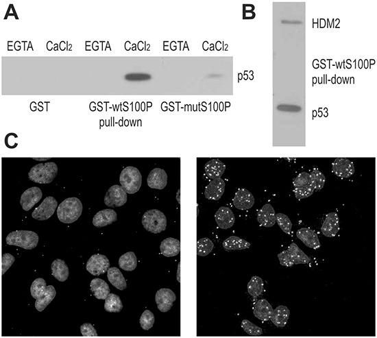
S100P belongs to the S100 family of calcium-binding proteins regulating diverse cellular processes. Certain S100 family members (S100A4 and S100B) are associated with cancer and used as biomarkers of metastatic phenotype. Also S100P is abnormally expressed in tumors and implicated in migration-invasion, survival, and response to therapy. Here we show that S100P binds the tumor suppressor protein p53 as well as its negative regulator HDM2, and that this interaction perturbs the p53-HDM2 binding and increases the p53 level. Paradoxically, the S100P-induced p53 is unable to activate its transcriptional targets hdm2, p21WAF, and bax following the DNA damage. This appears to be related to reduced phosphorylation of serine residues in both N-terminal and C-terminal regions of the p53 molecule. Furthermore, the S100P expression results in lower levels of pro-apoptotic proteins, in reduced cell death response to cytotoxic treatments, followed by stimulation of therapy-induced senescence and increased clonogenic survival. Conversely, the S100P silencing suppresses the ability of cancer cells to survive the DNA damage and form colonies. Thus, we propose that the oncogenic role of S100P involves binding and inactivation of p53, which leads to aberrant DNA damage responses linked with senescence and escape to proliferation. Thereby, the S100P protein may contribute to the outgrowth of aggressive tumor cells resistant to cytotoxic therapy and promote cancer progression.

Keywords: HDM2; S100P calcium-binding protein; cell death; p53 tumor suppressor; therapy-induced senescence.


Conflict of interest statement

The authors declare no conflict of interest.

Figures

Figure 1
Figure 1. S100P Interacts with p53 and HDM2
A. Interaction between S100P and p53 is demonstrated by GST-pulldown from T47D cells followed by the immunoblotting with the p53-specific antibody DO-1. The blot shows that the interaction is calcium-dependent and can be diminished by the F15A mutation compromising the dimerization of S100P. B. GST-pulldown from the RKO cells followed by immunoblotting reveals that S100P can bind both p53 (detected by the DO-1 antibody) and HDM2 (detected by the 2A9 antibody). C. Proximity ligation assay of MCF7 cells with endogenous S100P expression (control in left panel and treated with dexamethasone and UV irradiation in right panel) allowed for visualization of S100P-p53 interaction in situ. The PLA signal represented by the white spots shows stronger and more abundant interactions in treated cells with induced expression of S100P and p53.
Figure 2
Figure 2. S100P perturbs the p53-HDM2 interaction
The RKO cells were subjected to PLA analysis using the p53-specific rabbit polyclonal antibody CM1 and the HDM2-specific mouse monoclonal antibody 2A9. Panel A. shows the PLA signal for p53-HDM2 interaction in the mock-transfected cells under basal conditions, whereas panel B. shows the same cells after the treatment with UV irradiation, in which the signal is considerably elevated. Panels C. and D. show the S100P-transfected cells that exhibit reduced PLA signal under both basal and UV-treated conditions presumably due to S100P-perturbed interaction between p53 and HDM2.
Figure 3
Figure 3. S100P affects the expression of p53 and its transcriptional targets
A. Immunoblotting analysis of the p53 protein levels in relationship to S100P expression in A549 cells. The transiently transfected cells expressing S100P showed higher p53 protein level in both basal and treated conditions. B. Immunoblotting analysis of p53 and HDM2 protein levels in the mock-transfected and S100P-transfected RKO cells. RKO-S100P cells express higher levels of p53 and HDM2, but the level of HDM2 was not increased in response to treatment. UV = UV irradiation, PTX = paclitaxel, ETP = etoposide. C. Q-PCR analysis of p53 and its targets in the non-treated S100P vs mock-transfected cells shows increased levels of transcripts in the presence of S100P, D. Transcriptional analysis of p53 and its target genes in the mock-transfected RKO cells. Data show induction of the p53 mRNA itself as well as of the p53 protein targets in response to treatments. E. Transcriptional analysis by Q-PCR in S100P-expressing cells showed reduced levels of the analyzed transcripts in the cells subjected to treatments.
Figure 4
Figure 4. S100P influences the expression of cell death-associated proteins and improves cell viability
A. Protein expression was analyzed using the proteome-profiler array in extracts from the mock-transfected, camptothecin-treated (6h) vs untreated cells and in the transiently S100P-transfected, treated vs. untreated cells. Proteins showing remarkable changes are indicated by arrows and named at one of four corresponding panels. B. Graphical illustration of the changes in the p53 phosphorylation. All S100P expressing cells consistently showed decreased levels of phospho-serines upon treatment with different drugs (PTX=paclitaxel, ETP=etoposide, CPT=camptothecin). C. Graphical illustration of the cell viability following the drug treatment (determined by the propidium iodide and fluorescein diacetate staining of intact (non-fixed cells), left panel, and by the DNA labeling with propidium iodide in fixed cells, right panel). S100P-expressing cells (stable transfected mixed populations) showed significantly (*) increased viability compared to mock-transfected controls.
Figure 5
Figure 5. S100P induces the senescence-like morphology
A. Impedance-based real-time measurement of cell proliferation and/or death. Impedance values from quadruplicates are expressed as Cell index. B. Slopes derived from the same measurement data indicate the speed of changes in the cell numbers and/or cell-covered areas. C. Morphology of cells 72 h post-treatment with PTX, with the subset of S100P-expressing cells showing the senescence-like phenotype with flattened, granular appearance and visibly enlarged size (arrows). D. Immunostaining of p53 (red) and S100P (green), combined with the nuclear staining (blue) 72 h post-treatment with PTX. S100P and p53-positive cells display typical senescent morphology and contain abnormally large nuclei. Bottom right inlet reveals the p53 expression in the DAPI-stained nuclei after suppression of the S100P signal from the confocal image.
Figure 6
Figure 6. S100P contributes to therapy-induced senescence and survival
A. Detection of senescence by SA-β-galactosidase assay. Blue senescent cells were more frequent in PTX and ETP-treated S100P expressing RKO cells compared to mock controls, whereas no difference between these cell variants is visible under basal non-treated conditions. B. Representative image of colonies formed from the S100P-overexpressing RKO cells and mock control cells surviving the CPT treatment.
Figure 7
Figure 7. Suppression of endogenous S100P reduces senescence and survival, and supports the proposed mechanism of the S100P contribution to cancer progression
A. Q-PCR analysis of the S100P-silenced versus scrambled MCF-7 cells showed increased levels of the p53 and p21WAF transcripts in response to treatment with PTV and UV, respectively, in contrast to the results in the treated S100P-overexpressing versus control RKO cells as shown above on Figure 3E. B. S100P-silenced MCF-7 cells also showed weaker SA-β-gal staining compared to scrambled control. C. Representative image of colonies formed from the S100P-deficient MCF-7 cells and scrambled control cells surviving the PTX treatment. D. Schematic illustration of the proposed mechanism of S100P action in cancer cells exposed to DNA damage. In the wild-type p53-competent tumor cells, cytotoxic treatment triggers the DNA damage response that induces the accumulation of the p53 protein and its increased transactivation activity, which then leads preferably to cell death. In the presence of S100P, the wild-type p53 levels increase as a result of the S100P-p53 interaction, but the transactivation capacity is lowered. This suboptimal p53 activation is apparently not sufficient to induce full death response and the cells tend to enter the senescence program as evidenced by their morphology and expression profile. Such S100P-promoted shift in the phenotype of the subset of tumor cells with the wild type p53 can potentially lead to cancer progression through senescence escape and therapy resistance.

Similar articles

Cited by

References

    1. Chen H, Xu C, Jin Q, Liu Z. S100 protein family in human cancer. Am J Cancer Res. 2014;4:89–115. - PMC - PubMed
    1. Shen Z, Deng H, Fang Y, Zhu X, Ye G, Yan L, Liu H, Li G. Identification of the interplay between SOX9 and S100P in the metastasis and invasion of colon carcinoma. Oncotarget. 2015;6:20672–84. doi: 10.18632/oncotarget.3967. - DOI - PMC - PubMed
    1. Hsu YL, Hung JY, Liang YY, Lin YS, Tsai MJ, Chou SH, Lu CY, Kuo PL. S100P interacts with integrin α7 and increases cancer cell migration and invasion in lung cancer. Oncotarget. 2015;6:29585–98. doi: 10.18632/oncotarget.4987. - DOI - PMC - PubMed
    1. Bertram J, Palfner K, Hiddemann W, Kneba M. Elevated expression of S100P, CAPL and MAGE 3 in doxorubicin-resistant cell lines: comparison of mRNA differential display reverse transcription-polymerase chain reaction and subtractive suppressive hybridization for the analysis of differential gene expression. Anticancer Drugs. 1998;9:311–17. - PubMed
    1. Arumugam T, Simeone DM, Van Golen K, Logsdon CD. S100P promotes pancreatic cancer growth, survival, and invasion. Clin Cancer Res. 2005;11:5356–64. - PubMed

MeSH terms