. 2016 Apr 12;7(15):20621-35.
doi: 10.18632/oncotarget.7992.

Loss of SOCS3 in myeloid cells prolongs survival in a syngeneic model of glioma

Affiliations

Loss of SOCS3 in myeloid cells prolongs survival in a syngeneic model of glioma

Braden C McFarland et al. Oncotarget. .

Abstract

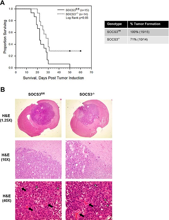
In glioma, microglia and macrophages are the largest population of tumor-infiltrating cells, referred to as glioma associated macrophages (GAMs). Herein, we sought to determine the role of Suppressor of Cytokine Signaling 3 (SOCS3), a negative regulator of Signal Transducer and Activator of Transcription 3 (STAT3), in GAM functionality in glioma. We utilized a conditional model in which SOCS3 deletion is restricted to the myeloid cell population. We found that SOCS3-deficient bone marrow-derived macrophages display enhanced and prolonged expression of pro-inflammatory M1 cytokines when exposed to glioma tumor cell conditioned medium in vitro. Moreover, we found that deletion of SOCS3 in the myeloid cell population delays intracranial tumor growth and increases survival of mice bearing orthotopic glioma tumors in vivo. Although intracranial tumors from mice with SOCS3-deficient myeloid cells appear histologically similar to control mice, we observed that loss of SOCS3 in myeloid cells results in decreased M2 polarized macrophage infiltration in the tumors. Furthermore, loss of SOCS3 in myeloid cells results in increased CD8+ T-cell and decreased regulatory T-cell infiltration in the tumors. These findings demonstrate a beneficial effect of M1 polarized macrophages on suppressing glioma tumor growth, and highlight the importance of immune cells in the tumor microenvironment.

Keywords: GL261; JAK/STAT; SOCS3; glioblastoma; macrophage.


Conflict of interest statement

CONFLICTS OF INTEREST

The authors declare no financial or competing conflicts of interests.

Figures

Figure 1
Figure 1. SOCS3-deficient macrophages exhibit prolonged activation of STAT3 when exposed to GL261 conditioned medium
(A and B) SOCS3fl/fl and SOCS3−/− BMDM were harvested from the femurs of 7–8 week old mice and cultured in RPMI 1640 containing 10% FBS and 10 ng/ml murine M-CSF for 5–7 days to expand. Cells were plated and at 24 h treated with GCM (50% volume) for the indicated times. Cells were lysed and immunoblotted with the indicated Abs (A) or RNA was isolated, cDNA generated and qRT-PCR performed for the indicated genes (B) Densitometric analysis is displayed as percent increase of SOCS3−/− compared to control SOCS3fl/fl macrophages. For example, at 1 h the SOCS3−/− macrophages display 30% higher p-STAT3 than the corresponding SOCS3fl/fl 1 h time point. *p < 0.05. Data are shown as mean ± S.D.
Figure 2
Figure 2. SOCS3−/− macrophages display enhanced M1 gene expression when exposed to GL261 conditioned medium
(AE) SOCS3fl/fl and SOCS3−/− BMDM were harvested from the femurs of 7–8 week old mice and cultured in RPMI 1640 containing 10% FBS and 10 ng/ml murine M-CSF for 5–7 days to expand. Cells were plated and at 24 h treated with GL261 conditioned medium (50% volume) for the indicated times. RNA was isolated, cDNA generated and qRT-PCR performed for the indicated genes. *p < 0.05. Data are shown as mean ± S.D.
Figure 3
Figure 3. Deletion of SOCS3 in myeloid cells prolongs in vivo survival
(A) SOCS3fl/fl and SOCS3−/− mice were injected intracranially with GL261 cells (1 × 10⁶ cells/5 μl). Mice were monitored for survival and euthanized at moribund. Combination of two independent experiments are shown. Log Rank p < 0.05. (B) At death, (times varied as shown in survival curve) brains from SOCS3fl/fl and SOCS3−/− mice injected with GL261 cells were formalin fixed, paraffin embedded, sectioned and H & E stained. Representative images are shown from each group. Black arrows indicate blood vessels and white arrow heads indicate mitotic figures.
Figure 4
Figure 4. GAM infiltration and morphological activation is similar between SOCS3fl/fl and SOCS3−/− tumor bearing mice
SOCS3fl/fl and SOCS3−/− mice were injected intracranially with GL261 cells (1 × 10⁶ cells/5 μl). Mice were monitored for survival and euthanized at moribund. Brains were formalin fixed, paraffin embedded, sectioned and stained with Iba1. Representative images are shown from each group. Top and middle panels, 10× and 40× Iba1 staining of brains with both tumor and normal brain shown. Black dotted line denotes border between tumor (left) and normal brain (right). Bottom panels, 40× Iba1 staining of intracranial tumor section.
Figure 5
Figure 5. Loss of SOCS3 in myeloid cells does not affect the quantity of myeloid cell tumor infiltration
(A–F) SOCS3fl/fl (n = 4) and SOCS3−/− (n = 4) mice were injected intracranially with GL261 cells (1 × 10⁶ cells/5 μl). Mice were euthanized on day 15, and mononuclear cells were isolated from the brains/tumors and analyzed by flow cytometry for macrophages (CD11b+ F4/80+ CD45hi), microglia (CD11b+ F4/80+ CD45mid), monocytes (CD11bmid Gr-1mid), or neutrophils (CD11bhi Gr-1hi), and were gated and quantified. Data are shown as mean ± S.D.
Figure 6
Figure 6. SOCS3−/− mice with intracranial tumors have diminished numbers of infiltrating Arg1+ M2 polarized cells compared to SOCS3fl/fl mice
SOCS3fl/fl mice (n = 4) and SOCS3−/− mice (n = 4) were injected with GL261-Luc cells (1 × 10⁶ cells/5 μl). Mice were euthanized on day 14, and mononuclear cells were isolated from the brains/tumors and analyzed by flow cytometry for Arg1 expression (CD45+ CD11b+ Arg1+) and iNOS expression (CD45+ CD11b+ iNOS+). *p < 0.05. Data are shown as mean ± S.D.
Figure 7
Figure 7. Loss of SOCS3 in myeloid cells results in increased CD8+ T-cell and decreased Treg infiltration in tumors
(A and C) SOCS3fl/fl (n = 11) and SOCS3−/− (n = 10) mice were injected intracranially with GL261-Luc cells (1 × 10⁶ cells/5 μl). Mice were euthanized on days 13–14 for analysis of infiltrating CD8+ T-cells (CD8+) and CD4+ T-cells (CD4+) in the brains and quantified by flow cytometry. Combination of two experiments are shown. *p < 0.05. (B) SOCS3fl/fl (n = 9) and SOCS3−/− (n = 9) mice were injected intracranially with GL261-Luc cells (1 × 10⁶ cells/5 μl). Mice were euthanized on days 13 or 21 for analysis of infiltrating Tregs (CD4+CD25+Foxp3+). Combination of two experiments are shown. *p < 0.05. Data are shown as mean ± S.D. (D and E) Representative histograms of CD4+, CD8+ and Treg populations are shown for each genotype.

References

    1. Dunn GP, Rinne ML, Wykosky J, Genovese G, Quayle SN, Dunn IF, Agarwalla PK, Chheda MG, Campos B, Wang A, Brennan C, Ligon KL, Furnari F, et al. Emerging insights into the molecular and cellular basis of glioblastoma. Genes Dev. 2012;26:756–784. - PMC - PubMed
    1. Li W, Graeber MB. The molecular profile of microglia under the influence of glioma. Neuro-Oncol. 2012;14:958–978. - PMC - PubMed
    1. Benveniste EN, Liu Y, McFarland BC, Qin H. Involvement of the janus kinase/signal transducer and activator of transcription signaling pathway in multiple sclerosis and the animal model of experimental autoimmune encephalomyelitis. J Interferon Cytokine Res. 2014;34:577–588. - PMC - PubMed
    1. Biswas SK, Allavena P, Mantovani A. Tumor-associated macrophages: functional diversity, clinical significance, and open questions. Semin Immunopathol. 2013;35:585–600. - PubMed
    1. Umemura N, Saio M, Suwa T, Kitoh Y, Bai J, Nonaka K, Ouyang GF, Okada M, Balazs M, Adany R, Shibata T, Takami T. Tumor-infiltrating myeloid-derived suppressor cells are pleiotropic-inflamed monocytes/macrophages that bear M1- and M2-type characteristics. J Leukoc Biol. 2008;83:1136–1144. - PubMed

MeSH terms

Substances