

MEK1 signaling promotes self-renewal and tumorigenicity of liver cancer stem cells via maintaining SIRT1 protein stabilization
- PMID: 26967560
- PMCID: PMC4991478
- DOI: 10.18632/oncotarget.7972
MEK1 signaling promotes self-renewal and tumorigenicity of liver cancer stem cells via maintaining SIRT1 protein stabilization
Abstract
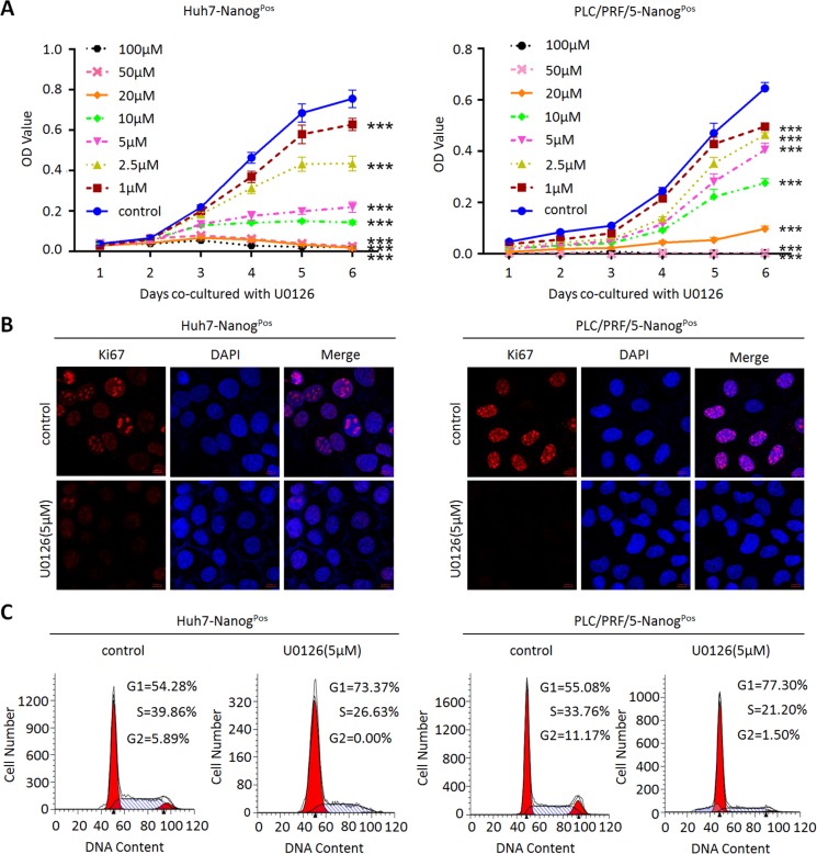
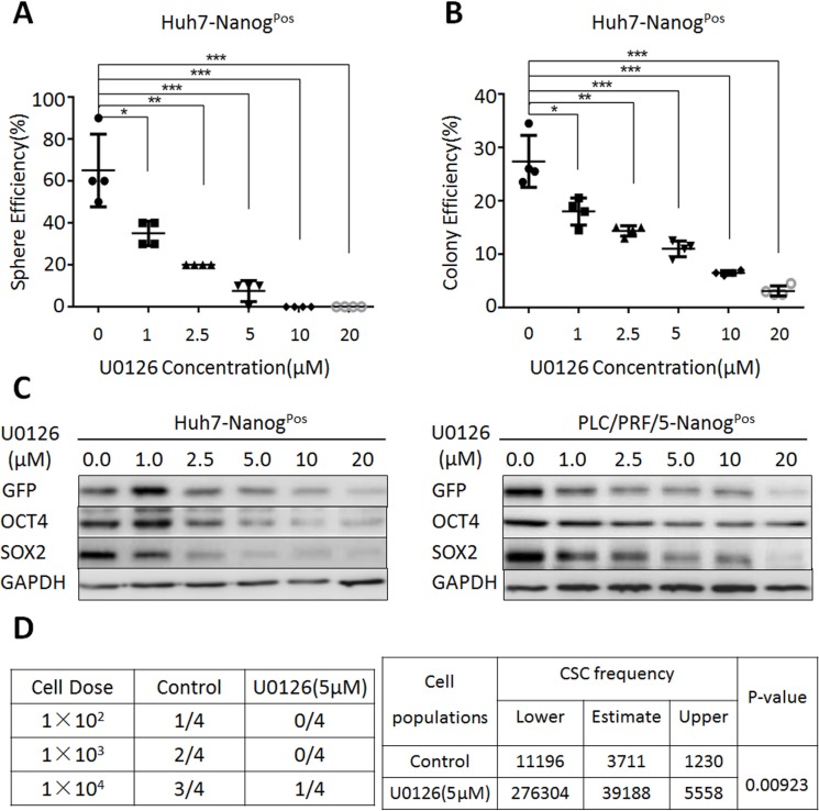
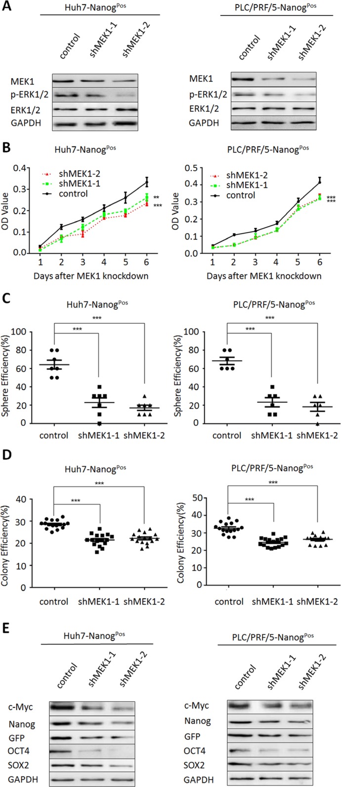
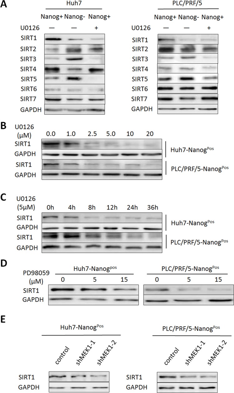
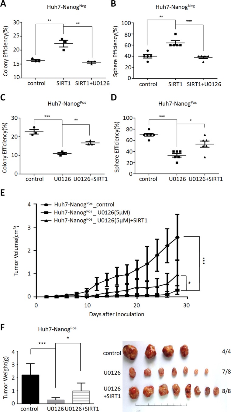
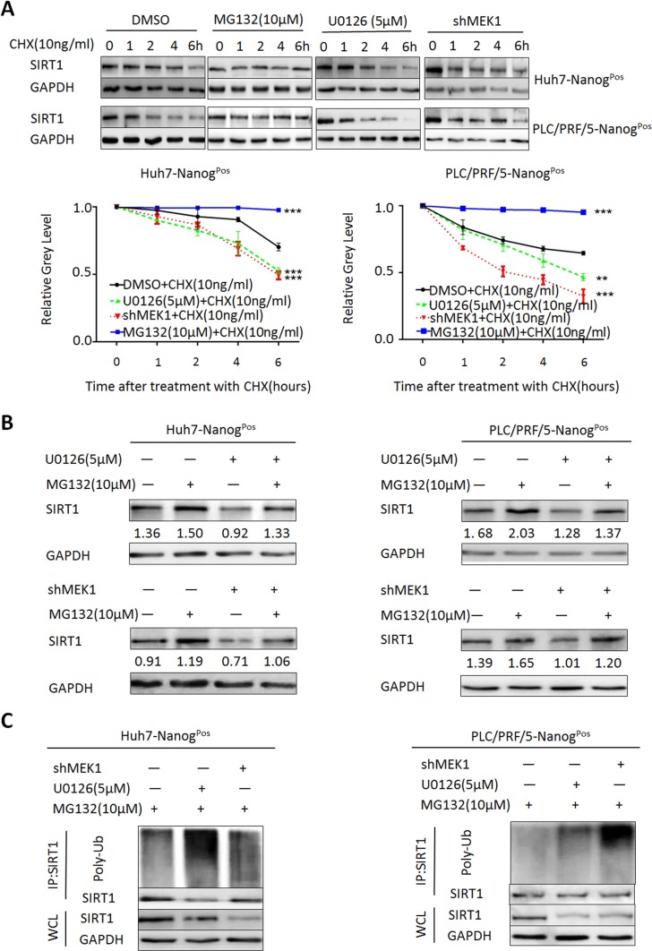
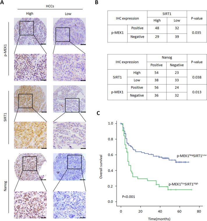
Hepatocellular carcinoma (HCC) is the third leading cause of cancer death. This high mortality has been commonly attributed to the presence of residual cancer stem cells (CSCs). Meanwhile, MEK1 signaling is regarded as a key molecular in HCC maintenance and development. However, nobody has figured out the particular mechanisms that how MEK1 signaling regulates liver CSCs self-renewal. In this study, we show that inhibition or depletion of MEK1 can significantly decrease liver CSCs self-renewal and tumor growth both in vitro and vivo conditions. Furthermore, we demonstrate that MEK1 signaling promotes liver CSCs self-renewal and tumorigenicity by maintaining SIRT1 level. Mechanistically, MEK1 signaling keeps SIRT1 protein stabilization through activating SIRT1 ubiquitination, which inhibits proteasomal degradation. Clinical analysis shows that patients co-expression of MEK1 and SIRT1 are associated with poor survival. Our finding indicates that MEK1-SIRT1 can act as a novel diagnostic biomarker and inhibition of MEK1 may be a viable therapeutic option for targeting liver CSCs treatment.
Keywords: MEK1 signaling; SIRT1; cancer stem cells (CSCs); hepatocellular carcinoma (HCC); proteasome degradation.
Conflict of interest statement
No potential conflicts of interest were disclosed.
Figures
Similar articles
-
SIRT1-mediated transcriptional regulation of SOX2 is important for self-renewal of liver cancer stem cells.Hepatology. 2016 Sep;64(3):814-27. doi: 10.1002/hep.28690. Epub 2016 Jul 18. Hepatology. 2016. PMID: 27312708
-
lncARSR promotes liver cancer stem cells expansion via STAT3 pathway.Gene. 2019 Mar 1;687:73-81. doi: 10.1016/j.gene.2018.10.087. Epub 2018 Oct 31. Gene. 2019. PMID: 30391438
-
Sox9 regulates self-renewal and tumorigenicity by promoting symmetrical cell division of cancer stem cells in hepatocellular carcinoma.Hepatology. 2016 Jul;64(1):117-29. doi: 10.1002/hep.28509. Epub 2016 Mar 25. Hepatology. 2016. PMID: 26910875
-
Role of stem cells in normal liver and cancer.Anticancer Agents Med Chem. 2011 Jul;11(6):522-8. doi: 10.2174/187152011796011091. Anticancer Agents Med Chem. 2011. PMID: 21554200 Review.
-
Noncoding RNAs in liver cancer stem cells: The big impact of little things.Cancer Lett. 2018 Apr 1;418:51-63. doi: 10.1016/j.canlet.2018.01.001. Epub 2018 Jan 4. Cancer Lett. 2018. PMID: 29307614 Review.
Cited by
-
Prognostic and clinicopathologic significance of SIRT1 expression in hepatocellular carcinoma.Oncotarget. 2016 Dec 22;8(32):52357-52365. doi: 10.18632/oncotarget.14096. eCollection 2017 Aug 8. Oncotarget. 2016. PMID: 28881735 Free PMC article.
-
Wnt/β-Catenin signaling pathway in hepatocellular carcinoma: pathogenic role and therapeutic target.Front Oncol. 2024 Apr 2;14:1367364. doi: 10.3389/fonc.2024.1367364. eCollection 2024. Front Oncol. 2024. PMID: 38634048 Free PMC article. Review.
-
Defect of SIRT1-FoxO3a axis is associated with the production of reactive oxygen species during protein kinase CK2 downregulation-mediated cellular senescence and nematode aging.BMB Rep. 2019 Apr;52(4):265-270. doi: 10.5483/BMBRep.2019.52.4.156. BMB Rep. 2019. PMID: 30103847 Free PMC article.
-
Targeting liver cancer stem cells for the treatment of hepatocellular carcinoma.Therap Adv Gastroenterol. 2019 Jan 22;12:1756284818821560. doi: 10.1177/1756284818821560. eCollection 2019. Therap Adv Gastroenterol. 2019. PMID: 30719075 Free PMC article. Review.
-
Sirt1 deacetylates and stabilizes p62 to promote hepato-carcinogenesis.Cell Death Dis. 2021 Apr 14;12(4):405. doi: 10.1038/s41419-021-03666-z. Cell Death Dis. 2021. PMID: 33854041 Free PMC article.
References
-
- El-Serag HB, Rudolph KL. Hepatocellular carcinoma: epidemiology and molecular carcinogenesis. Gastroenterology. 2007;132:2557–2576. - PubMed
-
- Zheng R, Zeng H, Zhang S, Chen T, Chen W. National estimates of cancer prevalence in China, 2011. Cancer Lett. 2016;370:33–38. - PubMed
-
- DeSantis CE, Lin CC, Mariotto AB, Siegel RL, Stein KD, Kramer JL, Alteri R, Robbins AS, Jemal A. Cancer treatment and survivorship statistics, 2014. CA Cancer J Clin. 2014;64:252–271. - PubMed
-
- Aguayo A, Patt YZ. Nonsurgical treatment of hepatocellular carcinoma. Semin Oncol. 2001;28:503–513. - PubMed
-
- Llovet JM, Bruix J. Systematic review of randomized trials for unresectable hepatocellular carcinoma: Chemoembolization improves survival. Hepatology. 2003;37:429–442. - PubMed
MeSH terms
Substances
LinkOut - more resources
Full Text Sources
Other Literature Sources
Medical
Miscellaneous
