. 2016 Apr;186(4):912-26.
doi: 10.1016/j.ajpath.2015.12.004. Epub 2016 Mar 8.

Epigallocatechin-3-Gallate Inhibition of Myeloperoxidase and Its Counter-Regulation by Dietary Iron and Lipocalin 2 in Murine Model of Gut Inflammation

Affiliations

Epigallocatechin-3-Gallate Inhibition of Myeloperoxidase and Its Counter-Regulation by Dietary Iron and Lipocalin 2 in Murine Model of Gut Inflammation

Beng San Yeoh et al. Am J Pathol. 2016 Apr.

Abstract

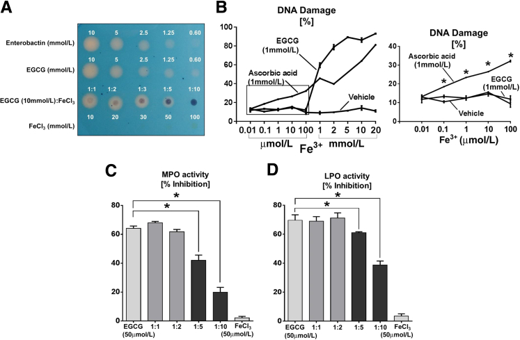
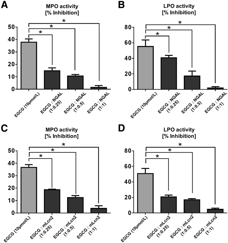
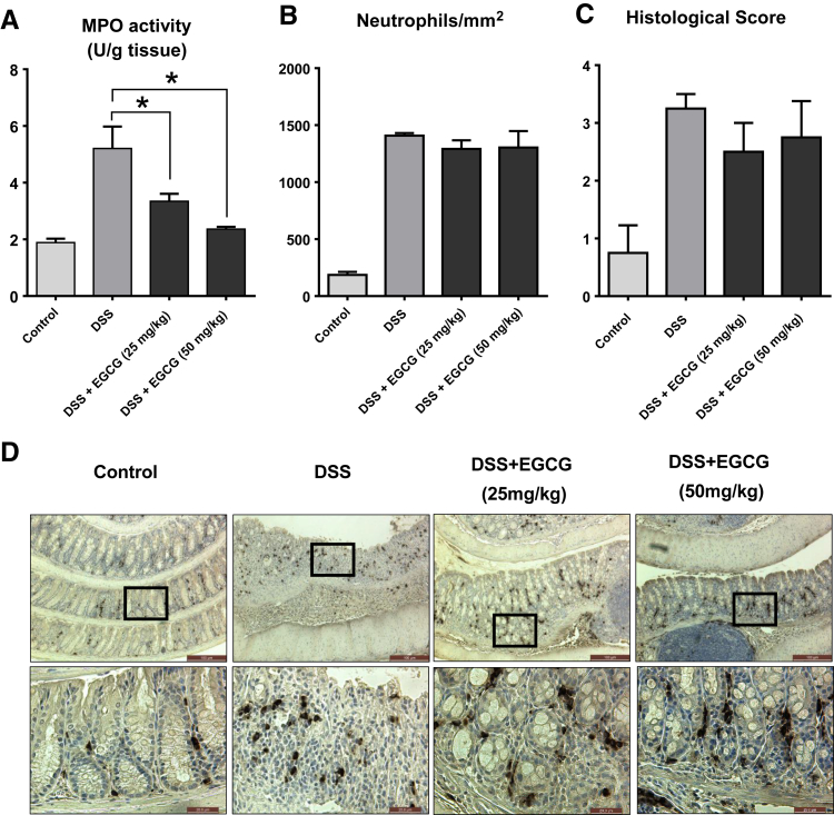
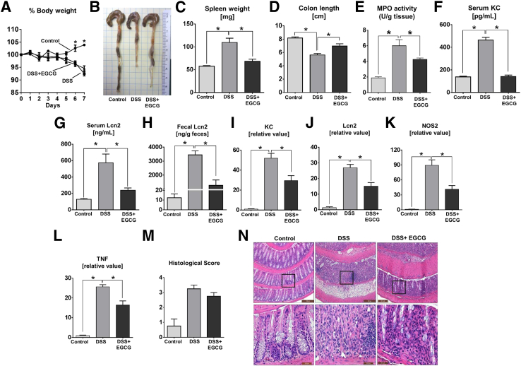
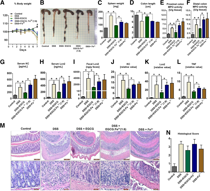
Green tea-derived polyphenol (-)-epigallocatechin-3-gallate (EGCG) has been extensively studied for its antioxidant and anti-inflammatory properties in models of inflammatory bowel disease, yet the underlying molecular mechanism is not completely understood. Herein, we demonstrate that EGCG can potently inhibit the proinflammatory enzyme myeloperoxidase in vitro in a dose-dependent manner over a range of physiologic temperatures and pH values. The ability of EGCG to mediate its inhibitory activity is counter-regulated by the presence of iron and lipocalin 2. Spectral analysis indicated that EGCG prevents the peroxidase-catalyzed reaction by reverting the reactive peroxidase heme (compound I:oxoiron) back to its native inactive ferric state, possibly via the exchange of electrons. Further, administration of EGCG to dextran sodium sulfate-induced colitic mice significantly reduced the colonic myeloperoxidase activity and alleviated proinflammatory mediators associated with gut inflammation. However, the efficacy of EGCG against gut inflammation is diminished when orally coadministered with iron. These findings indicate that the ability of EGCG to inhibit myeloperoxidase activity is one of the mechanisms by which it exerts mucoprotective effects and that counter-regulatory factors such as dietary iron and luminal lipocalin 2 should be taken into consideration for optimizing clinical management strategies for inflammatory bowel disease with the use of EGCG treatment.


Figures

Figure 1
Figure 1
EGCG inhibits and LPO activity. A and B: EGCG or ABAH was preincubated at the indicated final concentrations with either 12.5 μg/mL MPO or 25 μg/mL LPO for 5 minutes in 20-μL reaction before adding 30 μL of 2.2 mmol/L H2O2 and 50 μL of 100 mmol/L guaiacol. Line graph represents EGCG dose-dependent inhibition of MPO (A) and LPO (B) activity. C: Line graph represents inhibition of MPO activity by 50 μmol/L EGCG or 50 μmol/L ABAH which was added at indicated time points after MPO activity was initiated. For the in vitro bacterial killing assay, indicated final concentrations of EGCG were preincubated with 5 μg/mL MPO for 5 minutes, and then 30 μmol/L H2O2 was added to the mixture. K12 Escherichia coli (2 × 106 CFU/mL) was added to the reaction mixture and incubated for 30 minutes at room temperature. D: Percentage of bacteria killing by MPO was based on bacteria that were serially diluted from the reaction, plated on nonselective Lysogeny broth plates, and quantified as bacterial colony-forming units after overnight incubation at 37°C. Results are expressed as means ± SEM. Assays were performed in triplicates and are representative of three independent experiments. P < 0.05. ABAH, 4-aminobenzoic acid hydrazide; EGCG, (−)-epigallocatechin-3-gallate; H2O2, hydrogen peroxide; LPO, lactoperoxidase; MPO, myeloperoxidase.
Figure 2
Figure 2
Ferric iron-bound EGCG loses its ability to inhibit MPO and LPO activity. A: EGCG was preincubated with or without Fe3+ at indicated molar ratio and then 1 μL of the mixture was placed on CAS plate. Formation of orange halo indicates that EGCG chelated the iron from the CAS plate. Ent and FeCl3 were used as positive and negative control, respectively. In the bleomycin-detectable Fe2+ assay, EGCG or ascorbic acid (positive control) was incubated with indicated doses of Fe3+ in the reaction mixture for 1 hour at 37°C. B: Line graphs represent the conversion of Fe3+ to Fe2+, which was quantitated as Fe2+-dependent bleomycin-induced DNA damage. C and D: Bar graphs represent inhibition of MPO (C) and LPO (D) activity by 50 μmol/L EGCG that was preincubated with Fe3+ for 5 minutes at the indicated molar ratio before the assay was initiated. Results are expressed as means ± SEM. Assays were performed in a 96-well plate in triplicates and are representative of three independent experiments. P < 0.05. CAS, chrome Azurol S; EGCG, (−)-epigallocatechin-3-gallate; Ent, enterobactin; Fe2+, free ferrous iron; Fe3+, ferric iron; FeCl3 iron(III) chloride; LPO, lactoperoxidase; MPO, myeloperoxidase.
Figure 3
Figure 3
Human NGAL and its murine ortholog Lcn2 protect MPO and LPO from EGCG-mediated inhibition. EGCG (10 μmol/L) and indicated molar ratio of NGAL/Lcn2 were preincubated with either 12.5 μg/mL MPO or 25 μg/mL LPO for 5 minutes before the assay was initiated. A and B: Bar graphs represent inhibition of MPO (A) and LPO (B) activity by EGCG + NGAL at indicated molar ratio. C and D: Bar graph represents inhibition of MPO (C) and LPO (D) activity by EGCG preincubated with murine Lcn2 at indicated molar ratio. Results are expressed as means ± SEM. Assays were performed in a 96-well plate in triplicates and are representative of three independent experiments. P < 0.05. EGCG, (−)-epigallocatechin-3-gallate; Lcn2, lipocalin 2; LPO, lactoperoxidase; MPO, myeloperoxidase; NGAL, neutrophil gelatinase-associated lipocalin.
Figure 4
Figure 4
EGCG prevents the formation of peroxidase-catalyzed hypohalous acid. To perform spectral analysis, 1 mg/mL LPO was incubated with either 50 μmol/L EGCG or 50 μmol/L ABAH for 5 minutes. Reaction was initiated at room temperature by adding 30 μmol/L H2O2. Spectra were recorded at 250 to 500 nm with each spectrum representing an average of six scans taken in 1.0 second. AF: Image represents LPO alone (A), LPO + EGCG (B), LPO + ABAH (C), LPO + H2O2 (D), LPO + EGCG + H2O2 (E), and LPO + ABAH + H2O2 (F). Arrows indicate the direction of spectral changes over time on the initiation of the reaction. Results are representative of three independent experiments. ABAH, 4-aminobenzoic acid hydrazide; EGCG, (−)-epigallocatechin-3-gallate; H2O2, hydrogen peroxide; LPO, lactoperoxidase.
Figure 5
Figure 5
EGCG inhibits colonic MPO activity in the inflamed gut of colitic mice. DSS-induced colitic mice were orally gavaged with 25 or 50 mg EGCG/kg bodyweight at 24 and 3 hours before euthanasia. Colons were collected and processed for MPO assay. AC: Bar graph represents the colonic MPO activity (A), neutrophil count (B), and colon histologic score (C). D: Images represent immunohistochemistry staining for neutrophil marker Ly6G. Results are expressed as means ± SEM. n = 4 DSS-induced colitic mice. P < 0.05. Original magnification: ×100 (D, upper row); ×400 (D, lower row, enlarged from corresponding boxed areas above.). DSS, dextran sodium sulfate; EGCG, (−)-epigallocatechin-3-gallate; MPO, myeloperoxidase.
Figure 6
Figure 6
EGCG attenuates DSS-induced colitis in mice. DSS-induced colitic mice were orally gavaged with daily dose of either phosphate-buffered saline (vehicle) or 5 mg EGCG/kg body weight for 7 days and then euthanized 3 hours after the last EGCG dose. AL: The following variables were analyzed: body weight (A), gross colon (B), spleen weight (C), colon length (D), colonic myeloperoxidase activity (E), serum KC (F), serum Lcn2 (G), and fecal Lcn2 (H). Quantitative real-time RT-PCR analysis was used to quantify mRNA expression of KC (I), Lcn2 (J), NOS2 (K), and TNF (L). mRNA values are represented as fold-change normalized to 36B4 housekeeping gene. M and N: Colon histologic score (M) and hematoxylin and eosin staining of colon Swiss rolls at 100× (upper panel) and 400× (lower panel) magnification (N). Results are expressed as means ± SEM. n = 4 DSS-induced colitic mice. P < 0.05. Original magnification: ×100 (N, upper row); ×400 (N, lower row, enlarged from the boxed areas in the corresponding panels above). DSS, dextran sodium sulfate; EGCG, (−)-epigallocatechin-3-gallate; KC, keratinocyte-derived chemokine CXCL1; Lcn2, lipocalin 2; NOS2, nitric oxide synthase 2; TNF, tumor necrosis factor.
Figure 7
Figure 7
Oral administration of iron-saturated ECGC fails to mediate MPO inhibitory activity in vivo in mice with DSS-induced acute colitis. DSS-induced colitic mice were orally gavaged on day 6 and 7 (24 and 3 hours before euthanasia, respectively) with either phosphate-buffered saline (vehicle), 50 mg/kg body weight EGCG alone, or EGCG + iron (50 mg/kg bodyweight with iron co-administered at EGCG:iron molar ratio of 1:6). Control mice received only regular drinking water. AI: The following variables were analyzed: body weight (A), gross colon (B), spleen weight (C), colon length (D), MPO activity at the proximal colon (E) and distal colon (F), serum KC (G), serum Lcn2 (H), and fecal Lcn2 (I). Quantitative real-time RT-PCR analysis was used to quantify mRNA expression of KC (J), Lcn2 (K), and TNF (L). mRNA values are represented as fold-change normalized to 36B4 housekeeping gene. M and N: Hematoxylin and eosin staining of colon Swiss rolls (M) and their corresponding histologic score (N). Results expressed as means ± SEM. n = 4 DSS-induced colitic mice. P < 0.05. Original magnification: ×100× (M, upper row); ×400 (M, lower row). DSS, dextran sodium sulfate; EGCG, (−)-epigallocatechin-3-gallate; KC, keratinocyte-derived chemokine CXCL1; Lcn2, lipocalin 2; MPO, myeloperoxidase; TNF, tumor necrosis factor.
Figure 8
Figure 8
Potential mechanism by which EGCG mediates antioxidant effects that could alleviate gut inflammation. During inflammation, neutrophils release a plethora of pro-oxidant enzymes; one such enzyme is MPO whose bioactivity is associated with flares of inflammatory bowel disease. EGCG potently inhibits the activity of MPO and therefore reduces the level of oxidative stress in the inflamed gut. However, the beneficial MPO inhibition by EGCG is counter-regulated by iron and host Lcn2/NGAL. Altogether, the present study unravels the complex regulation of EGCG on MPO activity and its counter-regulation by iron and host Lcn2/NGAL. EGCG, (−)-epigallocatechin-3-gallate; Fe3+, ferric iron; Lcn2, lipocalin 2; MPO, myeloperoxidase; NGAL, neutrophil gelatinase-associated lipocalin.

Similar articles

Cited by

References

    1. Kim H.S., Quon M.J., Kim J.A. New insights into the mechanisms of polyphenols beyond antioxidant properties; lessons from the green tea polyphenol, epigallocatechin 3-gallate. Redox Biol. 2014;2:187–195. - PMC - PubMed
    1. Bose M., Lambert J.D., Ju J., Reuhl K.R., Shapses S.A., Yang C.S. The major green tea polyphenol, (−)-epigallocatechin-3-gallate, inhibits obesity, metabolic syndrome, and fatty liver disease in high-fat-fed mice. J Nutr. 2008;138:1677–1683. - PMC - PubMed
    1. Oz H.S., Chen T., de Villiers W.J. Green tea polyphenols and sulfasalazine have parallel anti-inflammatory properties in colitis models. Front Immunol. 2013;4:132. - PMC - PubMed
    1. Steinmann J., Buer J., Pietschmann T., Steinmann E. Anti-infective properties of epigallocatechin-3-gallate (EGCG), a component of green tea. Br J Pharmacol. 2013;168:1059–1073. - PMC - PubMed
    1. Dragicevic N., Smith A., Lin X., Yuan F., Copes N., Delic V., Tan J., Cao C., Shytle R.D., Bradshaw P.C. Green tea epigallocatechin-3-gallate (EGCG) and other flavonoids reduce Alzheimer's amyloid-induced mitochondrial dysfunction. J Alzheimers Dis. 2011;26:507–521. - PubMed

Publication types