. 2016 Jun;15(3):446-54.
doi: 10.1111/acel.12448. Epub 2016 Mar 10.

P66SHC deletion improves fertility and progeric phenotype of late-generation TERC-deficient mice but not their short lifespan

Affiliations

P66SHC deletion improves fertility and progeric phenotype of late-generation TERC-deficient mice but not their short lifespan

Marco Giorgio et al. Aging Cell. 2016 Jun.

Abstract

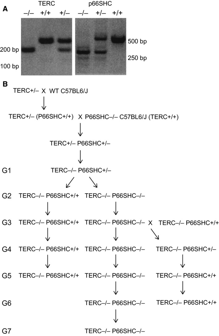
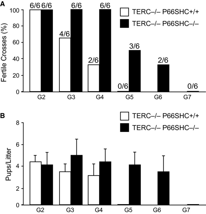
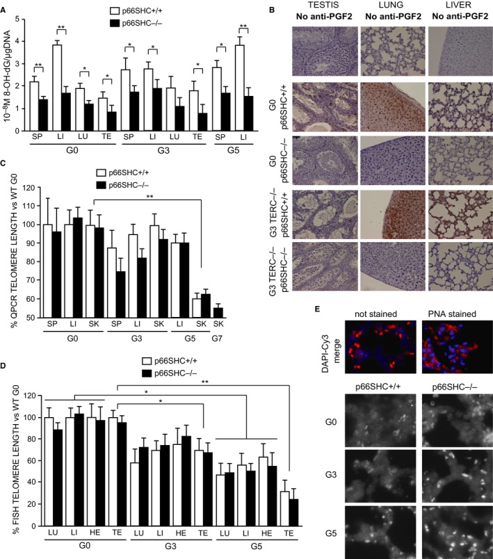
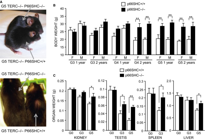
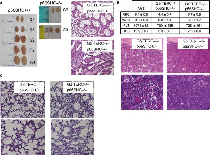
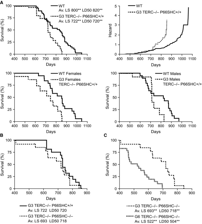
Oxidative stress and telomere attrition are considered the driving factors of aging. As oxidative damage to telomeric DNA favors the erosion of chromosome ends and, in turn, telomere shortening increases the sensitivity to pro-oxidants, these two factors may trigger a detrimental vicious cycle. To check whether limiting oxidative stress slows down telomere shortening and related progeria, we have investigated the effect of p66SHC deletion, which has been shown to reduce oxidative stress and mitochondrial apoptosis, on late-generation TERC (telomerase RNA component)-deficient mice having short telomeres and reduced lifespan. Double mutant (TERC(-/-) p66SHC(-/-) ) mice were generated, and their telomere length, fertility, and lifespan investigated in different generations. Results revealed that p66SHC deletion partially rescues sterility and weight loss, as well as organ atrophy, of TERC-deficient mice, but not their short lifespan and telomere erosion. Therefore, our data suggest that p66SHC-mediated oxidative stress and telomere shortening synergize in some tissues (including testes) to accelerate aging; however, early mortality of late-generation mice seems to be independent of any link between p66SHC-mediated oxidative stress and telomere attrition.

Keywords: fertility; lifespan; oxidative DNA damage; telo-meres.


Figures

Figure 1
Figure 1
Genealogy of TERC −/− p66SHC +/+ and TERC −/− p66SHC −/− mice. (A) PCR genotype analysis for TERC (left panel) and p66SHC mutation (right panel). (B) Scheme of the crosses performed to generate the different generations of TERC −/− p66SHC +/+ and TERC −/− p66SHC −/− mice.
Figure 2
Figure 2
Fertility of different generations of TERC −/− p66SHC +/+ and TERC −/− p66SHC −/− mice. (A) Percentage of productive crosses along generations (n = 6 couples per genotype per generation). (B) Average number ±SD of pups per litter along generations.
Figure 3
Figure 3
Oxidative stress and telomere length in TERC −/− p66SHC +/+ and TERC −/− p66SHC −/− mice. (A) Levels of 8‐OHdG per μg of DNA in TERC −/− p66SHC +/+ (white bars) and TERC −/− p66SHC −/− (black bars) spleen (SP), liver (LI), lung (LU), and testis (TE) measured by competitive enzyme immunoassay. Average values ±SD from n = 5 individuals for the G0 group and n = 3 individuals for the G3 and G5 groups are reported: * for < 0.05 and ** for P < 0.01. (B) 8‐isoprostane IHC representative images of testis, lung, and liver from G0 and G3 TERC −/− p66SHC +/+ and TERC −/− p66SHC −/− mice. Level of background signal is shown in the first three unstained slices. (C and D) Telomere length (expressed as a percentage versus the telomere length of C57Bl/6J WT mice) in spleen (SP), liver (LI), lung (LU), heart (HE), testis (TE) and skin (SK) from TERC −/− p66SHC +/+ and TERC −/− p66SHC −/− mice of different generation as evaluated by qPCR (C) where the average values ±SD from n = 3 individuals per group, measured in quadruplicates, are reported or Q‐FISH (D) where the average values ±SD from n = 3 individuals per group, each sample evaluated from two different sections for a total of around 40 nuclei, are reported. * for P < 0.05, ** for P < 0.01. (E) Representative FISH images (Cy3 alone grayscale and DAPI‐Cy3 merged color) obtained from the lung of different generation TERC −/− p66SHC +/+ and TERC −/− p66SHC −/− mice.
Figure 4
Figure 4
Appearance and weight of TERC −/− p66SHC +/+ and TERC −/− p66SHC −/− mice. (A) Representative images of 3 months old G5 TERC −/− p66SHC +/+ and TERC −/− p66SHC −/− mice. (B) Mean body weight of TERC −/− p66SHC +/+ and TERC −/− p66SHC −/− mice at 1 and 2 years of age (F, female; M, male), *for p < 0.05 ** for P < 0.01. (C) Kidney, testis, spleen, and liver weight of G0–G5 TERC −/− p66SHC +/+ and TERC −/− p66SHC −/− mice.
Figure 5
Figure 5
Phenotypic characterization of TERC −/− p66SHC +/+ and TERC −/− p66SHC −/− mice. (A) Representative images of morphology (left) and histology (right) of testes from TERC −/− p66SHC +/+ and TERC −/− p66SHC −/− mice. (B) Blood counts (upper table: WBC 103/μL, RBC 106/μL, PLT 103/μL, HGB mg dL−1; n = 6 mice per group) and bone marrow histology of TERC −/− p66SHC +/+ and TERC −/− p66SHC −/− mice. (C) Histological examination of lung tissues from G5 TERC −/− p66SHC +/+ and TERC −/− p66SHC −/− mice.
Figure 6
Figure 6
Survival curves of WT, TERC −/− p66SHC +/+, and TERC −/− p66SHC −/− mice. (A) Left upper graph, WT (n = 40) and G3 TERC −/− p66SHC +/+ (n = 18) male and female mice (Av. LS, average lifespan; LD50, median life time; **P < 0.01 for the effect of TERC genotype); right upper graph, cumulative hazard function for WT and G3 TERC −/− p66SHC +/+ male and female mice only; left lower graph, WT (n = 20) and G3 TERC −/− p66SHC +/+ (n = 10) female mice; right lower graph, WT (n = 20) and G3 TERC −/− p66SHC +/+ (n = 8) male mice only. (B) G3 TERC −/− p66SHC +/+ (n = 18) and G3 TERC −/− p66SHC −/− (n = 14) male and female mice. (C) G3 TERC −/− p66SHC −/− (n = 14) and G6 TERC −/− p66SHC −/− (n = 22) male and female mice (Av. LS and LD50 **P < 0.01 for the effect of generation).

References

    1. Albiero M, Poncina N, Tjwa M, Ciciliot S, Menegazzo L, Ceolotto G, Vigili de Kreutzenberg S, Moura R, Giorgio M, Pelicci P, Avogaro A, Fadini GP (2014) Diabetes causes bone marrow autonomic neuropathy and impairs stem cell mobilization via dysregulated p66Shc and Sirt1. Diabetes 63, 1353–1365. - PubMed
    1. Berry A, Capone F, Giorgio M, Pelicci PG, de Kloet ER, Alleva E, Minghetti L, Cirulli F (2007) Deletion of the life span determinant p66Shc prevents age‐dependent increases in emotionality and pain sensitivity in mice. Exp. Gerontol. 422, 37–45. - PubMed
    1. Blackburn EH, Greider CW, Szostak JW (2006) Telomeres and telomerase: The path from maize, Tetrahymena and yeast to human cancer and aging. Nat. Med. 12, 1133–1138. - PubMed
    1. Carpi A, Menabò R, Kaludercic N, Pelicci P, Di Lisa F, Giorgio M (2009) The cardioprotective effects elicited by p66(Shc) ablation demonstrate the crucial role of mitochondrial ROS formation in ischemia/reperfusion injury. Biochim. Biophys. Acta 1787, 774–780. - PubMed
    1. Daniali L, Benetos A, Susser E, Kark JD, Labat C, Kimura M, Desai K, Granick M, Aviv A (2013) Telomeres shorten at equivalent rates in somatic tissues of adults. Nat. Commun. 4, 1597. - PMC - PubMed

Publication types