. 2016 Apr;38(2):38.
doi: 10.1007/s11357-016-9902-5. Epub 2016 Mar 11.

Differential effects of IGF-1 deficiency during the life span on structural and biomechanical properties in the tibia of aged mice

Affiliations

Differential effects of IGF-1 deficiency during the life span on structural and biomechanical properties in the tibia of aged mice

Nicole M Ashpole et al. Age (Dordr). 2016 Apr.

Abstract

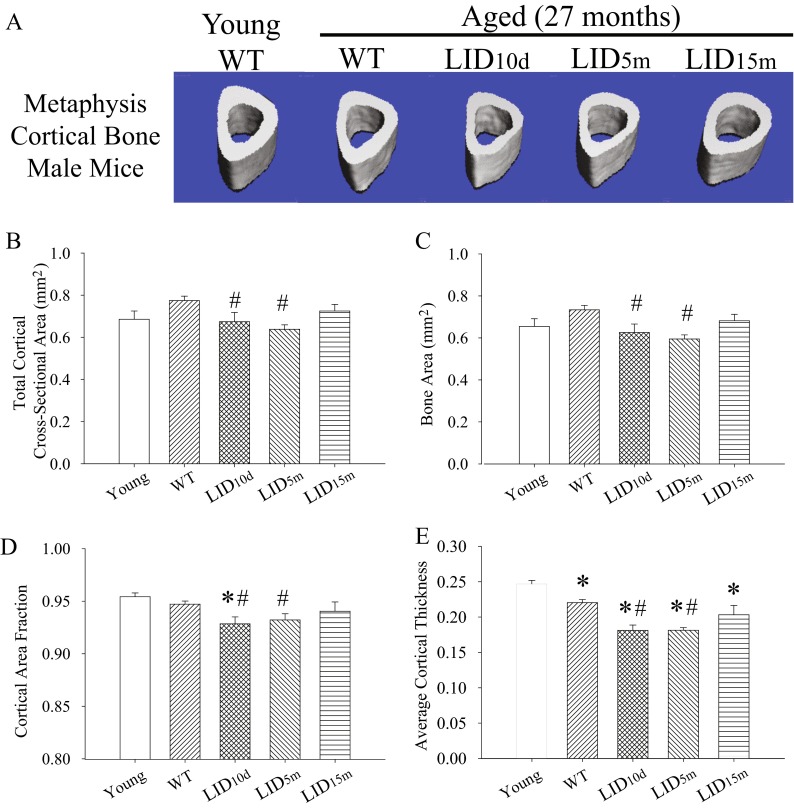
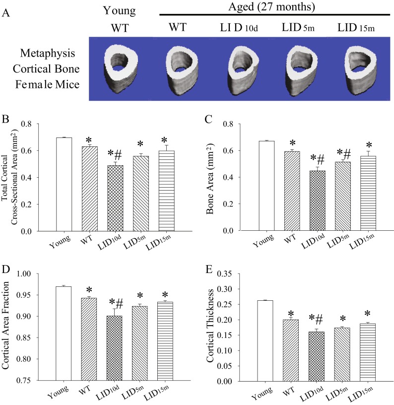
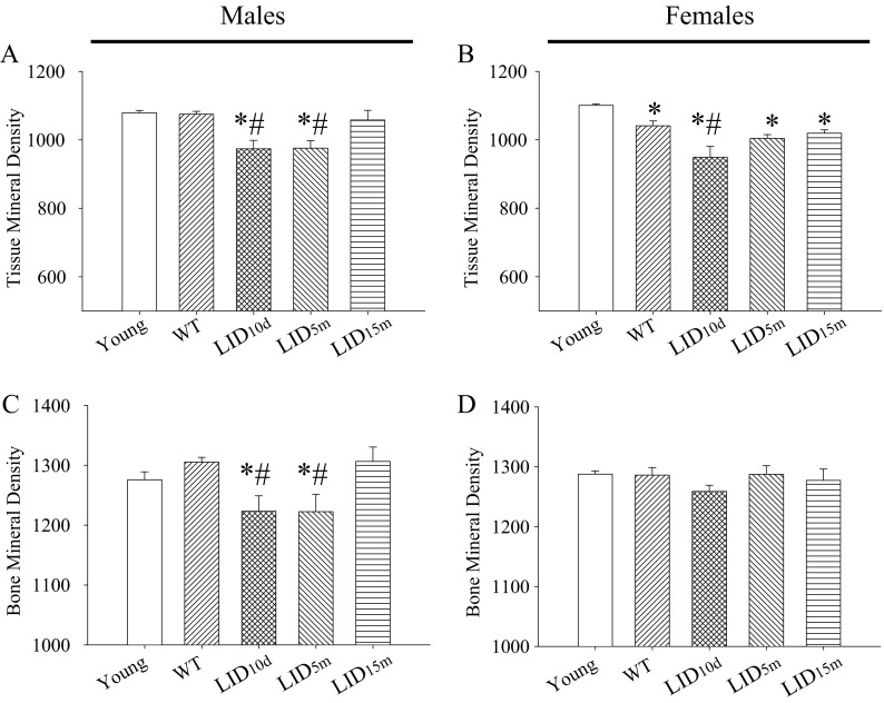
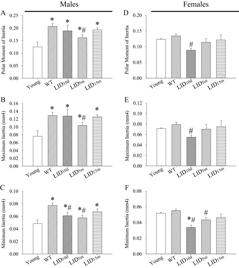
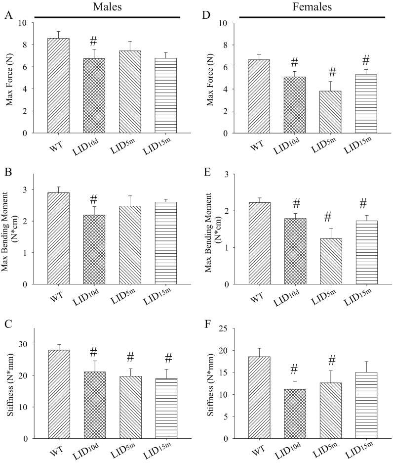
Advanced aging is associated with the loss of structural and biomechanical properties in bones, which increases the risk for bone fracture. Aging is also associated with reductions in circulating levels of the anabolic signaling hormone, insulin-like growth factor (IGF)-1. While the role of IGF-1 in bone development has been well characterized, the impact of the age-related loss of IGF-1 on bone aging remains controversial. Here, we describe the effects of reducing IGF-1 at multiple time points in the mouse life span--early in postnatal development, early adulthood, or late adulthood on tibia bone aging in both male and female igf (f/f) mice. Bone structure was analyzed at 27 months of age using microCT. We find that age-related reductions in cortical bone fraction, cortical thickness, and tissue mineral density were more pronounced when IGF-1 was reduced early in life and not in late adulthood. Three-point bone bending assays revealed that IGF-1 deficiency early in life resulted in reduced maximum force, maximum bending moment, and bone stiffness in aged males and females. The effects of IGF-1 on bone aging are microenvironment specific, as early-life loss of IGF-1 resulted in decreased cortical bone structure and strength along the diaphysis while significantly increasing trabecular bone fraction and trabecular number at the proximal metaphysis. The increases in trabecular bone were limited to males, as early-life loss of IGF-1 did not alter bone fraction or number in females. Together, our data suggest that the age-related loss of IGF-1 influences tibia bone aging in a sex-specific, microenvironment-specific, and time-dependent manner.

Keywords: IGF-1; MicroCT; Three-point bone bending; Tibia.


Conflict of interest statement

Compliance with ethical standards All procedures were approved by and followed the guidelines of the Institutional Animal Care and Use Committee and veterinarians at University of Oklahoma Health Sciences Center (OUHSC).

Figures

Fig. 1
Fig. 1
Early-life reductions in IGF-1 lead to decreased cortical bone structure in aged males. a Representative images of the cortical bone in the metaphysis of the tibia in young (n = 3), aged (n = 19), and aged IGF-1-deficient male mice (n = 6–7 per group). Average b cross-sectional area, c bone area, d cortical area fraction (BV/TV), and e cortical thickness in the metaphysis of the tibia in young, aged, and aged IGF-1-deficient male mice. The pound sign indicates a significant difference compared to wild-type controls at that time point, while the asterisk indicates a significant difference in the wild-type mice compared to the young 2-month time point, *#p < 0.05, mean ± SEM
Fig. 2
Fig. 2
Early-life reductions in IGF-1 lead to decreased cortical bone structure in aged females. a Representative images of the cortical bone in the metaphysis of the tibia in young (n = 3), aged (n = 18), and aged IGF-1-deficient female mice (n = 5–7). Average b cross-sectional area, c bone area, d cortical area fraction (BV/TV), and e cortical thickness in the metaphysis of the tibia in young, aged, and aged IGF-1-deficient female mice. The pound sign indicates a significant difference compared to wild-type controls at that time point, while the asterisk indicates a significant difference in the wild-type mice compared to the young 2-month time point, *#p < 0.05, mean ± SEM
Fig. 3
Fig. 3
Early-life reductions in IGF-1 lead to decreased mineral density in aged mice. Average tissue mineral density in young (n = 3), aged (n = 18–19), and aged IGF-1-deficient a male and b female mice (n = 5–7). Average tissue mineral density in young, aged, and aged IGF-1-deficient c male and d female mice. The pound sign indicates a significant difference compared to wild-type controls at that time point, while the asterisk indicates a significant difference in the wild-type mice compared to the young 2-month time point, *#p < 0.05, mean ± SEM
Fig. 4
Fig. 4
IGF-1 influences moments of inertia in the tibia of aged mice. Average a polar moment of inertia, b maximum inertia, and c minimum inertia in the tibia of young, aged, and aged IGF-1-deficient male mice. Average d polar moment of inertia, e maximum inertia, and f minimum inertia in the tibia of young, aged, and aged IGF-1-deficient male mice. The pound sign indicates a significant difference compared to wild-type controls at that time point, while the asterisk indicates a significant difference in the wild-type mice compared to the young 2-month time point, *#p < 0.05, mean ± SEM
Fig. 5
Fig. 5
Early-life reductions in IGF-1 result in decreased bone bending properties late in life. Average a force applied to the tibia before bone breaking, b maximum bending moment, and c stiffness in the tibia of wild-type (n = 12) and IGF-1-deficient male mice (n = 4–6 per group), as measured by three-point bending test. Average d force applied to the tibia before bone breaking, e maximum bending moment, and f stiffness in the tibia of wild-type (n = 10) and IGF-1-deficient female mice (n = 4–5 per group). The pound sign indicates a significant difference compared to wild-type controls at that time point, #p < 0.05, mean ± SEM

References

    1. Ashpole NM, et al. IGF-1 regulates vertebral bone aging through sex-specific and time-dependent mechanisms. J Bone Mineral Res: Off J Am Soc Bone Miner Res. 2015 - PMC - PubMed
    1. Baker J, Liu JP, Robertson EJ, Efstratiadis A. Role of insulin-like growth factors in embryonic and postnatal growth. Cell. 1993;75:73–82. doi: 10.1016/S0092-8674(05)80085-6. - DOI - PubMed
    1. Bando H, Zhang C, Takada Y, Yamasaki R, Saito S. Impaired secretion of growth hormone-releasing hormone, growth hormone and IGF-I in elderly men. Acta Endocrinol. 1991;124:31–36. - PubMed
    1. Behrendt AK, et al. Dietary restriction-induced alterations in bone phenotype: effects of lifelong versus short-term caloric restriction on femoral and vertebral bone in C57BL/6 mice. J Bone Miner Res: Off J Am Soc Bone Miner Res. 2015 - PubMed
    1. Bikle D, et al. The skeletal structure of insulin-like growth factor I-deficient mice. J Bone Miner Res off J Am Soc Bone Miner Res. 2001;16:2320–2329. doi: 10.1359/jbmr.2001.16.12.2320. - DOI - PubMed

Publication types

Substances

LinkOut - more resources