Review
. 2016 Oct;139 Suppl 2(Suppl Suppl 2):115-125.
doi: 10.1111/jnc.13486. Epub 2016 Mar 10.

Bioenergetics and redox adaptations of astrocytes to neuronal activity

Affiliations
Review

Bioenergetics and redox adaptations of astrocytes to neuronal activity

Juan P Bolaños. J Neurochem. 2016 Oct.

Abstract

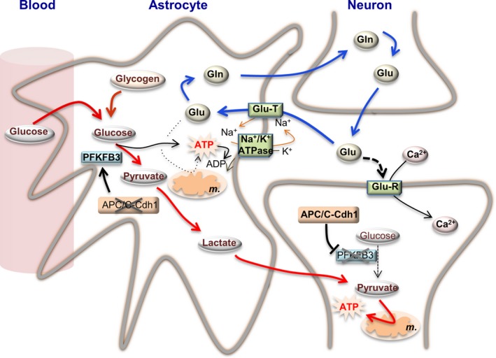
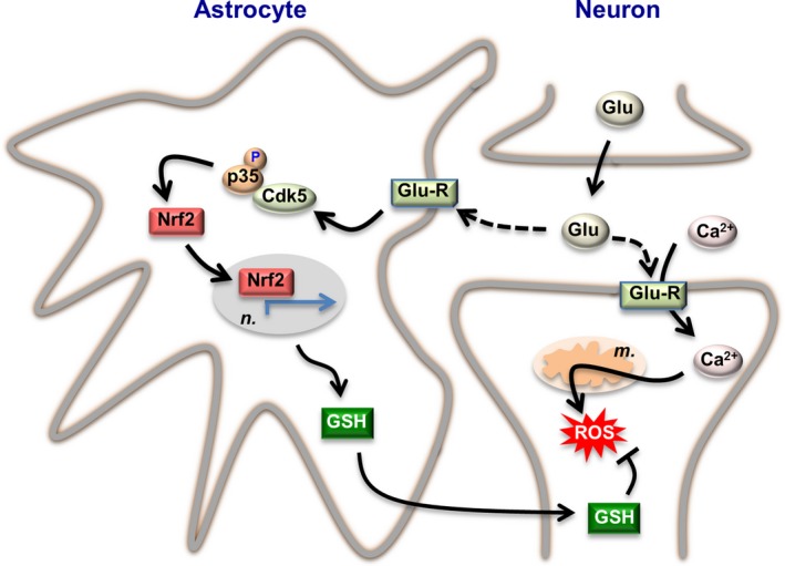
Neuronal activity is a high-energy demanding process recruiting all neural cells that adapt their metabolism to sustain the energy and redox balance of neurons. During neurotransmission, synaptic cleft glutamate activates its receptors in neurons and in astrocytes, before being taken up by astrocytes through energy costly transporters. In astrocytes, the energy requirement for glutamate influx is likely to be met by glycolysis. To enable this, astrocytes are constitutively glycolytic, robustly expressing 6-phosphofructo-2-kinase/fructose-2,6-bisphosphatase-3 (PFKFB3), an enzyme that is negligibly present in neurons by continuous degradation because of the ubiquitin-proteasome pathway via anaphase-promoting complex/cyclosome (APC)-Cdh1. Additional factors contributing to the glycolytic frame of astrocytes may include 5'-AMP-activated protein kinase (AMPK), hypoxia-inducible factor-1 (HIF-1), pyruvate kinase muscle isoform-2 (PKM2), pyruvate dehydrogenase kinase-4 (PDK4), lactate dehydrogenase-B, or monocarboxylate transporter-4 (MCT4). Neurotransmission-associated messengers, such as nitric oxide or ammonium, stimulate lactate release from astrocytes. Astrocyte-derived glycolytic lactate thus sustains the energy needs of neurons, which in contrast to astrocytes mainly rely on oxidative phosphorylation. Neuronal activity unavoidably triggers reactive oxygen species, but the antioxidant defense of neurons is weak; hence, they use glucose for oxidation through the pentose-phosphate pathway to preserve the redox status. Furthermore, neural activity is coupled with erythroid-derived erythroid-derived 2-like 2 (Nrf2) mediated transcriptional activation of antioxidant genes in astrocytes, which boost the de novo glutathione biosynthesis in neighbor neurons. Thus, the bioenergetics and redox programs of astrocytes are adapted to sustain neuronal activity and survival. Developing therapeutic strategies to interfere with these pathways may be useful to combat neurological diseases. Our current knowledge on brain's management of bioenergetics and redox requirements associated with neural activity is herein revisited. The astrocyte-neuronal lactate shuttle (ANLS) explains the energy needs of neurotransmission. Furthermore, neurotransmission unavoidably triggers increased mitochondrial reactive oxygen species in neurons. By coupling glutamatergic activity with transcriptional activation of antioxidant genes, astrocytes provide neurons with neuroprotective glutathione through an astrocyte-neuronal glutathione shuttle (ANGS). This article is part of the 60th Anniversary special issue.

Keywords: AMPK; GSH; Cdh1; Glycolysis; Nrf2; PFKFB3.


Figures

Figure 1
Figure 1
Bioenergetics adaptations of astrocytes to neurotransmission. Synaptic cleft glutamate (Glu) released by the pre‐synaptic neuron acts on glutamate receptors (Glu‐R) placed in the post‐synaptic neurons, triggering the influx of Ca2+ (and Na+, not indicated) and causing plasma membrane depolarization, which is needed to propagate the nervous impulse (neuronal activity). To reset basal levels of glutamate in the synaptic cleft, it is taken up by astrocytes through glutamate transporters (Glu‐T), which require Na+ uptake. Astrocytes convert glutamate into glutamine (Gln), which is released and then taken up by neurons, which convert it back into glutamate (blue arrowed lines). The Na+/K+ATPase hydrolyses ATP to conserve its energy to reset Na+ (and K+) homeostasis. Such ATP can be obtained by glutamate oxidation in the tricarboxylic acid cycle in the mitochondria (m.). However, strong evidence indicates that glutamate uptake is coupled, via a yet unknown mechanism, with glucose conversion into lactate through the glycolytic pathway (red arrowed lines). Neuronal activity is also coupled with glucose uptake from the blood, and with glycogen conversion into lactate. As neuronal activity‐associated glycolytic activation in astrocytes is coupled with stoichiometric lactate release, it is likely that glycolytic‐derived ATP would be in charge of sustaining Na+/K+ homeostasis during glutamate uptake. Astrocytes are genetically adapted to support a constitutive glycolytic phenotype in view of their low activity of APC/C‐Cdh1 (anaphase‐promoting complex/cyclosome/Cdh1), the E3 ubiquitin ligase responsible for the degradation of 6‐phosphofructo‐2‐kinase/fructose‐2,6‐bisphosphatase‐3 (PFKFB3), a pro‐glycolytic enzyme. In contrast to astrocytes, neurons express high APC/C‐Cdh1 activity hence continuously degrading PFKFB3 causing their low glycolytic phenotype. Thus, neurons need an energy source different from glucose. Evidence obtained from cultured cells and in vivo strongly suggest that astrocyte‐released lactate support neuronal functions, both serving as a fuel and, possibly, as a signaling molecule. Thus, astrocytes are metabolically adapted to sustain the bioenergetics status of neurons during neural activity. The stoichiometry of the reactions has been omitted for clarity. Likewise, additional factors involved in these adaptations could not be depicted herein and can be found in the main text.
Figure 2
Figure 2
Redox adaptation of astrocytes to neurotransmission. Synaptic cleft glutamate (Glu) released by the pre‐synaptic neuron acts on glutamate receptors (Glu‐R) placed both in the post‐synaptic neuron and astrocytes. Part of intracellular Ca2+ at the post‐synaptic neurons is removed from the cytosol by entering mitochondria, and this causes mitochondrial production of reactive oxygen species (ROS). Synaptic cleft glutamate interacts with its receptors placed in astrocytes, triggering a cascade of events via cyclin‐dependent kinase‐5 (Cdk5)‐mediated phosphorylation of Nrf2 (nuclear factor‐erythroid 2‐related factor‐2), which enters the nucleus (n.) and binds to the antioxidant responsive elements (ARE) to promote the expression of antioxidant genes (green arrowed lines). This pathway leads to the biosynthesis and release of glutathione (GSH), whose precursors are taken up by neurons for the de novo GSH biosynthesis necessary to detoxify neuronal activity‐mediated mitochondrial ROS. Thus, through this astrocyte‐neuronal glutathione shuttle, astrocytes sustain the redox status of neurons during neural activity. The stoichiometry of the reactions has been omitted for clarity. Likewise, additional factors involved in these adaptations could not be depicted herein and can be found in the main text.

References

    1. Allaman I., Belanger M. and Magistretti P. J. (2011) Astrocyte‐neuron metabolic relationships: for better and for worse. Trends Neurosci. 34, 76–87. - PubMed
    1. Allen N. J. and Barres B. A. (2009) Neuroscience: Glia ‐ more than just brain glue. Nature 457, 675–677. - PubMed
    1. Almeida A. and Bolaños J. P. (2001) A transient inhibition of mitochondrial ATP synthesis by nitric oxide synthase activation triggered apoptosis in primary cortical neurons. J. Neurochem. 77, 676–690. - PubMed
    1. Almeida A., Almeida J., Bolaños J. P. and Moncada S. (2001) Different responses of astrocytes and neurons to nitric oxide: the role of glycolytically‐generated ATP in astrocyte protection. Proc. Natl Acad. Sci. USA 98, 15294–15299. - PMC - PubMed
    1. Almeida A., Moncada S. and Bolaños J. P. (2004) Nitric oxide switches on glycolysis through the AMP protein kinase and 6‐phosphofructo‐2‐kinase pathway. Nat. Cell Biol. 6, 45–51. - PubMed

Publication types

Substances