Expression of forkhead box transcription factor genes Foxp1 and Foxp2 during jaw development
- PMID: 26969076
- PMCID: PMC4842334
- DOI: 10.1016/j.gep.2016.03.001
Expression of forkhead box transcription factor genes Foxp1 and Foxp2 during jaw development
Abstract
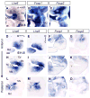
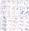
Development of the face is regulated by a large number of genes that are expressed in temporally and spatially specific patterns. While significant progress has been made on characterizing the genes that operate in the oral region of the face, those regulating development of the aboral (lateral) region remain largely unknown. Recently, we discovered that transcription factors LIM homeobox (LHX) 6 and LHX8, which are key regulators of oral development, repressed the expression of the genes encoding forkhead box transcription factors, Foxp1 and Foxp2, in the oral region. To gain insights into the potential role of the Foxp genes in region-specific development of the face, we examined their expression patterns in the first pharyngeal arch (primordium for the jaw) of mouse embryos at a high spatial and temporal resolution. Foxp1 and Foxp2 were preferentially expressed in the aboral and posterior parts of the first pharyngeal arch, including the developing temporomandibular joint. Through double immunofluorescence and double fluorescent RNA in situ hybridization, we found that Foxp1 was expressed in the progenitor cells for the muscle, bone, and connective tissue. Foxp2 was expressed in subsets of bone and connective tissue progenitors but not in the myoblasts. Neither gene was expressed in the dental mesenchyme nor in the oral half of the palatal shelf undergoing extensive growth and morphogenesis. Together, we demonstrated for the first time that Foxp1 and Foxp2 are expressed during craniofacial development. Our data suggest that the Foxp genes may regulate development of the aboral and posterior regions of the jaw.
Keywords: Craniofacial development; Foxp1; Foxp2; Mouse; Pharyngeal arch; Temporomandibular joint.
Copyright © 2016 Elsevier B.V. All rights reserved.
Figures


Similar articles
-
Foxp1 expression is essential for sex-specific murine neonatal ultrasonic vocalization.Hum Mol Genet. 2017 Apr 15;26(8):1511-1521. doi: 10.1093/hmg/ddx055. Hum Mol Genet. 2017. PMID: 28204507
-
Expression of Foxp4 in the developing and adult rat forebrain.J Neurosci Res. 2008 Nov 1;86(14):3106-16. doi: 10.1002/jnr.21770. J Neurosci Res. 2008. PMID: 18561326
-
Foxp2 and Foxp1 cooperatively regulate lung and esophagus development.Development. 2007 May;134(10):1991-2000. doi: 10.1242/dev.02846. Epub 2007 Apr 11. Development. 2007. PMID: 17428829
-
The distinct and overlapping phenotypic spectra of FOXP1 and FOXP2 in cognitive disorders.Hum Genet. 2012 Nov;131(11):1687-98. doi: 10.1007/s00439-012-1193-z. Epub 2012 Jun 27. Hum Genet. 2012. PMID: 22736078 Free PMC article. Review.
-
FOXP transcription factors in vertebrate brain development, function, and disorders.Wiley Interdiscip Rev Dev Biol. 2020 Sep;9(5):e375. doi: 10.1002/wdev.375. Epub 2020 Jan 30. Wiley Interdiscip Rev Dev Biol. 2020. PMID: 31999079 Free PMC article. Review.
Cited by
-
Association of low-frequency genetic variants in regulatory regions with nonsyndromic orofacial clefts.Am J Med Genet A. 2019 Mar;179(3):467-474. doi: 10.1002/ajmg.a.61002. Epub 2018 Dec 24. Am J Med Genet A. 2019. PMID: 30582786 Free PMC article.
-
The molecular anatomy of mammalian upper lip and primary palate fusion at single cell resolution.Development. 2019 Jun 17;146(12):dev174888. doi: 10.1242/dev.174888. Development. 2019. PMID: 31118233 Free PMC article.
-
Downregulation of SOX9 expression in developing entheses adjacent to intramembranous bone.PLoS One. 2024 May 10;19(5):e0301080. doi: 10.1371/journal.pone.0301080. eCollection 2024. PLoS One. 2024. PMID: 38728328 Free PMC article.
-
Targeted Sequencing of Candidate Regions Associated with Sagittal and Metopic Nonsyndromic Craniosynostosis.Genes (Basel). 2022 May 3;13(5):816. doi: 10.3390/genes13050816. Genes (Basel). 2022. PMID: 35627201 Free PMC article.
-
Dissecting human embryonic skeletal stem cell ontogeny by single-cell transcriptomic and functional analyses.Cell Res. 2021 Jul;31(7):742-757. doi: 10.1038/s41422-021-00467-z. Epub 2021 Jan 20. Cell Res. 2021. PMID: 33473154 Free PMC article.
References
-
- Cesario JM, Malt A Landin, Jeong J. Developmental Genetics of the Pharyngeal Arch System, e-book. In: Saint-Jeannet JP, Kessler D, editors. Colloquium Series on Developmental Biology. Vol. 6. Morgan & Claypool Life Sciences Publishers; 2015a.
Publication types
MeSH terms
Substances
Grants and funding
LinkOut - more resources
Full Text Sources
Other Literature Sources
Molecular Biology Databases
Research Materials

