. 2016 Jun;101(6):698-706.
doi: 10.3324/haematol.2015.137273. Epub 2016 Mar 11.

A distinct plasmablast and naïve B-cell phenotype in primary immune thrombocytopenia

Affiliations

A distinct plasmablast and naïve B-cell phenotype in primary immune thrombocytopenia

Shaun M Flint et al. Haematologica. 2016 Jun.

Abstract

Primary immune thrombocytopenia is an autoimmune disorder in which platelet destruction is a consequence of both B- and T-cell dysregulation. Flow cytometry was used to further characterize the B- and T-cell compartments in a cross-sectional cohort of 26 immune thrombocytopenia patients including antiplatelet antibody positive (n=14) and negative (n=12) patients exposed to a range of therapies, and a cohort of matched healthy volunteers. Markers for B-cell activating factor and its receptors, relevant B-cell activation markers (CD95 and CD21) and markers for CD4(+) T-cell subsets, including circulating T-follicular helper-like cells, were included. Our results indicate that an expanded population of CD95(+) naïve B cells correlated with disease activity in immune thrombocytopenia patients regardless of treatment status. A population of CD21-naïve B cells was specifically expanded in autoantibody-positive immune thrombocytopenia patients. Furthermore, the B-cell maturation antigen, a receptor for B-cell activating factor, was consistently and strongly up-regulated on plasmablasts from immune thrombocytopenia patients. These observations have parallels in other autoantibody-mediated diseases and suggest that loss of peripheral tolerance in naïve B cells may be an important component of immune thrombocytopenia pathogenesis. Moreover, the B-cell maturation antigen represents a potential target for plasma cell directed therapies in immune thrombocytopenia.


Figures

Figure 1.
Figure 1.
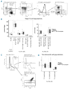
Analysis of major B-cell subsets: differences by diagnosis. (A) Gating strategy for B-cell subsets. (B) Major B-cell subsets expressed as a percentage of CD19+ cells and stratified by diagnosis (i.e. healthy volunteers or splenectomy- and rituximab-naïve immune thrombocytopenia patients). (C) Gating strategy for transitional B cells. Mitotracker positive gate defined using expression on switched memory B cells. Transitional B cells are Mitotracker+ IgD+CD27. T1 transitional population is CD10+; T2 and 3 populations are distinguished by CD38 expression. (D) Transitional B-cell populations, expressed as a percentage of B cells and stratified by diagnosis as in (B). Significant pairwise P-values (Wilcoxon signed-rank test) are shown. ritux/splen: rituximab or splenectomy; mem: memory. Plasmablasts are gated out of other B-cell subsets.
Figure 2.
Figure 2.
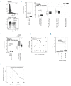
CD95+ (Fas receptor+) naïve B cells are more frequent in immune thrombocytopenia (ITP) patients. (A) Representative gating, showing distribution of CD95+ cells across the major B-cell subsets. (B) Proportions of CD95+ cells across B-cell subsets, stratified by diagnosis (i.e. healthy volunteers or splenectomy- and rituximab-naïve ITP patients). (C) Proportions of CD95+ naïve and IgD+CD27+ B cells stratified by diagnosis and prior rituximab or splenectomy. (D) Proportions of CD95+ naïve and IgD+CD27+ B cells by timing of rituximab, for those ITP patients who had received prior rituximab. (E) CD95+ cells as a proportion of naïve B cells, stratified by platelet count. (F) CD95+ naïve B cells by platelet count for ITP patients where a second time point is available (Δ Splenectomy; □ Rituximab; o Neither). Significant pairwise P-values (Wilcoxon signed-rank test) are shown in (B, C and E). (F) P-value is generated using a repeated measures linear mixed effects model. PB: plasmablast; DN: IgD-CD27- B cells; plt: platelet count; ritux/splen: rituximab or splenectomy. Plasmablasts are gated out of other B-cell subsets.
Figure 3.
Figure 3.
CD21 (complement receptor 2–) naïve B cells are more frequent in immune thrombocytopenia (ITP) patients. (A) Representative gating, showing distribution of CD21 cells across the major B-cell subsets. (B) Proportions of CD21– cells across B-cell subsets, stratified by diagnosis (i.e. healthy volunteers or splenectomy- and rituximab-naïve ITP patients). (C) CD21 cells as a proportion of naïve B cells, stratified by anti-platelet antibody status. Significant pairwise P-values (Wilcoxon signed-rank test) are shown. sw mem + PB: switched memory B cells and plasmablasts i.e. CD27+IgD B cells; DN: double-negative B cells, i.e. CD27IgD B cells; nonsw memory: non-switched memory, i.e. CD27+IgD+ B cells; Ab: antiplatelet antibody; ritux/splen: rituximab or splenectomy.
Figure 4.
Figure 4.
Analysis of serum BAFF and its receptors: BCMA is markedly up-regulated on the surface of plasmablasts in immune thrombocytopenia, regardless of activity or autoantibody status. (A) Serum BAFF, stratified by past treatment and diagnosis. BAFFR (B) and TACI (C) median fluorescence intensity (MFI) across the major B-cell subpopulations, stratified by diagnosis and prior treatment. (D) Plasmablast BCMA MFI, stratified by diagnosis and prior treatment. (E and F) Plasmablast BCMA MFI, stratified by platelet count and autoantibody status. Significant pairwise P-values (Wilcoxon signed-rank test) are shown. Plt: platelet count; ritux/splen: rituximab or splenectomy.
Figure 5.
Figure 5.
CD4+ T cells in immune thrombocytopenia (ITP) and healthy volunteer samples by diagnosis. (A) T-cell populations expressed as a proportion of their parent population, stratified by diagnosis [i.e. healthy volunteers (white, n=18) or splenectomy- and rituximab-naïve ITP patients (dark gray, n=9)]. (B) The same T-cell populations, expressed in absolute numbers [healthy volunteers (white, n=9); splenectomy- and rituximab-naïve ITP patients (dark gray, n=8)]. Significant pairwise P-values (Wilcoxon signed-rank test) are shown. ritux/splen: rituximab or splenectomy; mem: memory, i.e. CD45RA.

References

    1. Nørgaard M, Jensen A, Engebjerg MC, et al. Long-term clinical outcomes of patients with primary chronic immune thrombocytopenia: A Danish population-based cohort study. Blood. 2011;117(13):3514–3520. - PubMed
    1. Shulman NR, Marder VJ, Weinrach RS. Similarities between known antiplatelet antibodies and the factor responsible for thrombocytopenia in idiopathic purpura. Physiologic, serologic and isotopic studies. Ann N Y Acad Sci. 1965;124(2):499–542. - PubMed
    1. McMillan R. Antiplatelet Antibodies in Chronic Immune Thrombocytopenia and Their Role in Platelet Destruction and Defective Platelet Production. Hematol Oncol Clin North Am. 2009;23(6):1163–1175. - PubMed
    1. Zhu XJ, Shi Y, Peng J, et al. The effects of BAFF and BAFF-R-Fc fusion protein in immune thrombocytopenia. Blood. 2009;114(26):5362–5367. - PubMed
    1. Emmerich F, Bal G, Barakat A, et al. High-level serum B-cell activating factor and promoter polymorphisms in patients with idiopathic thrombocytopenic purpura. Br J Haematol. 2007;136(2):309–314. - PubMed

Publication types

MeSH terms