

Reflectance confocal microscopy features of mycosis fungoides and Sézary syndrome: correlation with histopathologic and T-cell receptor rearrangement studies
- PMID: 26969149
- PMCID: PMC5515474
- DOI: 10.1111/cup.12708
Reflectance confocal microscopy features of mycosis fungoides and Sézary syndrome: correlation with histopathologic and T-cell receptor rearrangement studies
Abstract
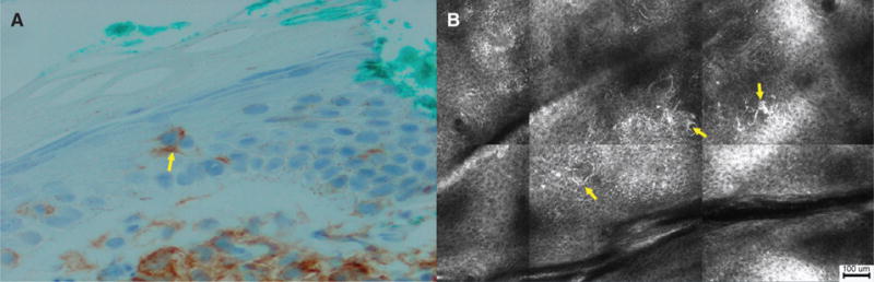
Background: Mycosis fungoides/Sézary syndrome (MF/SS) often requires multiple skin biopsies for definitive diagnosis. In vivo reflectance confocal microscopy (RCM) visualizes high-resolution cellular detail of the skin. The objective of this study is to evaluate the morphologic features of MF/SS using RCM and to correlate RCM features with histopathology and T-cell receptor (TCR) gene rearrangement studies.
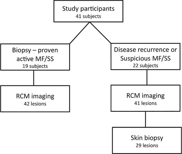
Methods: A cohort of patients with active/recurrent or suspicious MF/SS disease was prospectively recruited for RCM imaging and histopathologic/RCM images were evaluated. Statistical analyses were performed to identify unique RCM features and to correlate RCM features with histopathologic findings and TCR rearrangement studies.
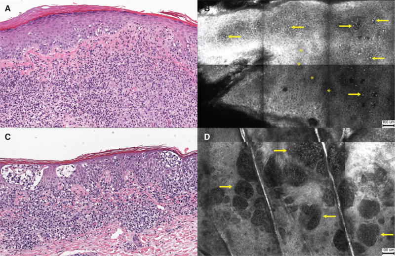
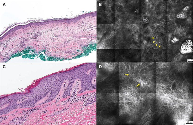
Results: Eighty-three lesions were evaluated. Correlation between RCM and histopathology was moderate for all relatable features (κ = 0.41, p<0.001), almost perfect for intraepidermal atypical lymphocytes [prevalence and bias-adjusted kappa (PABAK) = 0.90], and fair for Pautrier collections (κ = 0.32, p = 0.03). Lesions with Pautrier collections identified by RCM were significantly more likely to show TCR clonality (p = 0.04) and diagnostic features of MF/SS on histopathology (p = 0.03).
Conclusions: Our study captures morphologic RCM criteria for a variety of skin lesions. Pautrier collections visualized by RCM are associated with improved histopathologic diagnosis and detection of TCR gene clonality. Although further studies are needed to validate the diagnostic implications of RCM for MF/SS, our study highlights the potential utility of RCM.
Keywords: RCM; mycosis fungoides.
© 2016 John Wiley & Sons A/S. Published by John Wiley & Sons Ltd.
Figures

References
-
- Willemze R, Jaffe ES, Burg G, et al. WHO-EORTC classification for cutaneous lymphomas. Blood. 2005;105:3768. - PubMed
-
- Jawed SI, Myskowski PL, Horwitz S, Moskowitz A, Querfeld C. Primary cutaneous T-cell lymphoma (mycosis fungoides and Sezary syndrome): Part I. Diagnosis: clinical and histopathologic features and new molecular and biologic markers. J Am Acad Dermatol. 2014;70:205.e1. - PubMed
-
- Guitart J, Kennedy J, Ronan S, Chmiel JS, Hsiegh YC, Variakojis D. Histologic criteria for the diagnosis of mycosis fungoides: proposal for a grading system to standardize pathology reporting. J Cutan Pathol. 2001;28:174. - PubMed
-
- Agero AL, Gill M, Ardigo M, Myskowski P, Halpern AC, Gonzalez S. In vivo reflectance confocal microscopy of mycosis fungoides: a preliminary study. J Am Acad Dermatol. 2007;57:435. - PubMed
Publication types
MeSH terms
Substances
Grants and funding
LinkOut - more resources
Full Text Sources
Other Literature Sources
Medical
