. 2016 Jul;20(7):1307-18.
doi: 10.1111/jcmm.12812. Epub 2016 Mar 10.

Aspirin induces Nrf2-mediated transcriptional activation of haem oxygenase-1 in protection of human melanocytes from H2 O2 -induced oxidative stress

Affiliations

Aspirin induces Nrf2-mediated transcriptional activation of haem oxygenase-1 in protection of human melanocytes from H2 O2 -induced oxidative stress

Zhe Jian et al. J Cell Mol Med. 2016 Jul.

Abstract

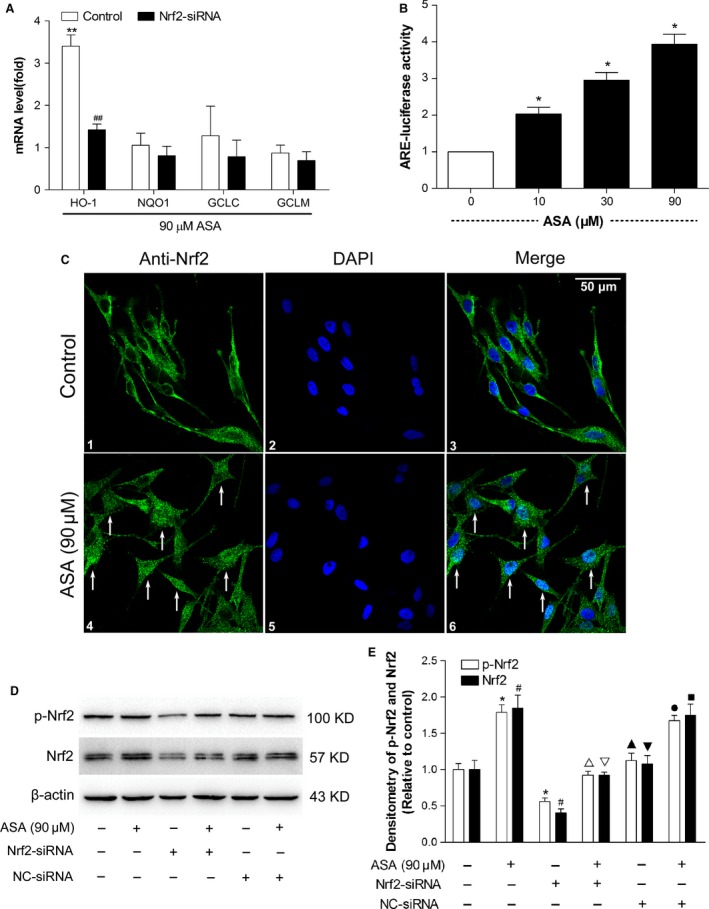
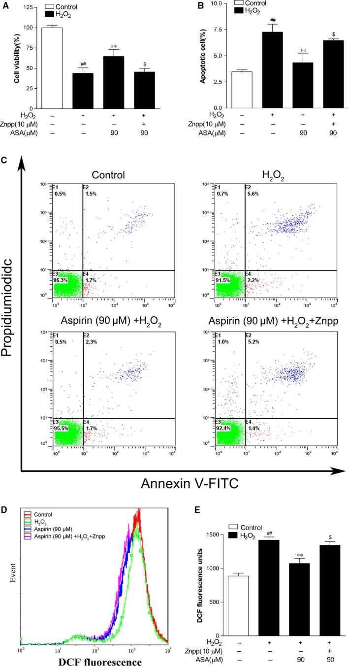
The removal of hydrogen peroxide (H2 O2 ) by antioxidants has been proven to be beneficial to patients with vitiligo. Aspirin (acetylsalicylic acid, ASA) has antioxidant activity and has great preventive and therapeutical effect in many oxidative stress-relevant diseases. Whether ASA can protect human melanocytes against oxidative stress needs to be further studied. Here, we investigated the potential protective effect and mechanisms of ASA against H2 O2 -induced oxidative injury in human melanocytes. Human melanocytes were pre-treated with different concentrations of ASA, followed by exposure to 1.0 mM H2 O2 . Cell apoptosis, intracellular reactive oxygen species (ROS) levels were evaluated by flow cytometry, and cell viability was determined by an Cell Counting Kit-8 assay. Total and phosphorylated NRF2 expression, NRF2 nuclear translocation and antioxidant response element (ARE) transcriptional activity were assayed with or without Nrf2-siRNA transfection to investigate the possible molecular mechanisms. Concomitant with an increase in viability, pre-treatment of 10-90 μmol/l ASA resulted in decreased rate of apoptotic cells, lactate dehydrogenase release and intracellular ROS levels in primary human melanocytes. Furthermore, we found ASA dramatically induced NRF2 nuclear translocation, enhanced ARE-luciferase activity, increased both p- NRF2 and total NRF2 levels, and induced the expression of haem oxygenase-1 (HO-1) in human melanocytes. In addition, knockdown of Nrf2 expression or pharmacological inhibition of HO-1 abrogated the protective action of ASA on melanocytes against H2 O2 -induced cytotoxicity and apoptosis. These results suggest that ASA protects human melanocytes against H2 O2 -induced oxidative stress via Nrf2-driven transcriptional activation of HO-1.

Keywords: aspirin; haem oxygenase-1; hydrogen peroxide; melanocyte; nuclear factor E2-related factor 2; oxidative stress; vitiligo.


Figures

Figure 1
Figure 1
Protective effect of aspirin on H2O2‐induced cytotoxicity in primary human melanocytes. (A) Primary human melanocytes were treated with different concentrations of aspirin for 1–5 days, and cell proliferation was determined by CCK‐8 assay. (B) The effect of aspirin on H2O2‐induced morphological changes in primary human melanocytes. (C) Primary human melanocytes were treated with various concentrations of aspirin alone for 24 hrs, and cell viability was determined by CCK‐8 assay. (D) Primary human melanocytes were exposed to various concentrations of aspirin (10, 30 and 90 μM) for 24 hrs. After pre‐treatment, cells were treated with 1.0 mM H2O2 for 24 hrs, and the cell viability was determined by CCK‐8 assay. Data are presented as the mean ± S.D. of the percentages observed in treated versus untreated control cells from three independent experiments. *P < 0.01 versus untreated control cells, **P < 0.01 versus H2O2‐treated cells, # P < 0.01 versus H2O2‐treated cells and ## P < 0.001 versus untreated control cells.
Figure 2
Figure 2
Effects of aspirin on LDH release and intracellular ROS levels in primary human melanocytes following H2O2 challenge. (A) LDH leakage of human melanocytes was determined by an LDH release assay. (B) Representative results for ROS production after pre‐treatment. (C) The fluorescence intensity of the cells was calculated relative to that of untreated control cells. The results shown in A and C are presented as the mean ± S.D. of three independent experiments. **P < 0.01 versus H2O2‐treated cells, # P < 0.05 and ## P < 0.01 versus untreated control cells.
Figure 3
Figure 3
Inhibitory effect of aspirin on H2O2‐induced apoptosis in primary human melanocytes. Cells were pre‐treated with or without aspirin at the indicated concentrations for 24 hrs and then incubated in the presence or absence of 1.0 mM H2O2 for a further 24 hrs. Cellular apoptosis was assayed by annexin V‐FITC and PI counterstaining and analyzed with flow cytometry. (A) The original representative flow cytometry figures. (B) The apoptosis rates of human melanocytes. The data are presented as the mean ± S.D. of three independent experiments. **P < 0.01 versus H2O2‐treated cells and ## P < 0.01 versus untreated control cells.
Figure 4
Figure 4
Investigation of Nrf2‐mediated phase II gene modulation, antioxidant response element (ARE) activity, Nrf2 nuclear translocation, NRF2 and p‐NRF2 protein levels in human melanocytes after treatment with aspirin. (A) Modulation of HO‐1, NQO‐1, GCLC and GCLM gene expression levels was detected by real‐time PCR in human melanocytes after a 24‐hr aspirin treatment with or without Nrf2‐siRNA or NC‐siRNA treatment. Data are shown as ratios of gene expression in treated cells to that in untreated controls after normalization on the basis of the expression of the GAPDH housekeeping gene. **P < 0.01 compared with the untreated cells. ## P < 0.01 compared with the control cells. (B) PIG1 cells were cotransfected with an ARE‐luciferase reporter plasmid (pGL3‐ARE) and a Renilla luciferase reporter plasmid (pRLtk) for 24 hrs and treated with aspirin (10, 30 and 90 μM) for 24 hrs before luciferase activity was measured. Firefly luciferase activity in relative light units per second (RLU/s) was normalized to Renilla luciferase activity and expressed as x‐fold multiples of the control to obtain a ratio of the experimental condition to the control (control cells without ASA treatment). *P < 0.05 compared with the control cells. (C) NRF2 localization in melanocytes was observed by laser confocal scanning microscopy after treatment with or without aspirin for 24 hrs. Nuclear NRF2 translocation and accumulation occurred in aspirin treatment group (Arrows indicated), scale bar = 50 μm. (D) NRF2 and p‐NRF2 protein levels were measured by western blots after treatment with 90 μM aspirin for 24 hrs. (E) the intensity of each band was quantified by densitometry analysis. All protein expression was normalized to that of β‐actin, which was used as an internal control. The data are presented as the mean ± S.D. of three independent experiments. *P < 0.05 compared with p‐NRF2 in control group (untreated). # P < 0.05 compared with NRF2 in control group (untreated). P < 0.05 compared with p‐NRF2 in only ASA‐treated group. P < 0.05 compared with NRF2 in only ASA‐treated group. Cropped gel images are used in this figure and the gels were run under the same experimental conditions. P > 0.05 compared with p‐NRF2 in control group (untreated). P > 0.05 compared with NRF2 in control group (untreated). P > 0.05 compared with p‐NRF2 in only ASA‐treated group. P > 0.05 compared with NRF2 in only ASA‐treated group.
Figure 5
Figure 5
Effect of Nrf2 siRNA and aspirin on cell viability, apoptosis and intracellular ROS level induced by H2O2 in PIG1 cells. (A) Viability of PIG1 cells pre‐treated with Nrf2 siRNA, NC‐siRNA and/or aspirin (ASA; 90 μM) was determined with CCK‐8 assay 24 hrs after exposure to 1.0 mM H2O2 (n = 3). (B) The apoptosis rates of PIG1 cells. (C) The effect of Nrf2 siRNA, NC‐siRNA and aspirin on H2O2‐induced morphological changes in PIG1 cells. (D) The original representative flow cytometry figures. (E) Representative results for ROS production after pre‐treatment. (F) The fluorescence intensity of the cells was calculated relative to that of untreated control cells. The data are presented as the mean ± S.D. of three independent experiments. **P < 0.01 and $ P > 0.05 versus H2O2‐treated cells; ## P < 0.01 versus untreated control cells. P > 0.05 versus H2O2+ ASA treated cells.
Figure 6
Figure 6
Effect of Znpp and aspirin on cell viability, apoptosis and intracellular ROS level induced by H2O2 in PIG1 cells. (A) Viability of PIG1 cells pre‐treated with Znpp and/or aspirin (ASA; 90 μM) was determined with CCK‐8 assay 24 hrs after exposure to 1.0 mM H2O2 (n = 3). (B) The apoptosis rates of PIG1 cells. (C) The original representative flow cytometry figures. (D) Representative results for ROS production after pre‐treatment. (E) The fluorescence intensity of the cells was calculated relative to that of untreated control cells. The data are presented as the mean ± S.D. of three independent experiments. **P < 0.01 and $ P > 0.05 versus H2O2‐treated cells; ## P < 0.01 versus untreated control cells.

References

    1. Halder RM, Chappell JL. Vitiligo update. Semin Cutan Med Surg. 2009; 28: 86–92. - PubMed
    1. Schallreuter KU, Bahadoran P, Picardo M, et al Vitiligo pathogenesis: autoimmune disease, genetic defect, excessive reactive oxygen species, calcium imbalance, or what else? Exp Dermatol. 2008; 17: 139–40, 141–60. - PubMed
    1. Picardo M. Bastonini. A new view of vitiligo: looking at normal‐appearing skin. J Invest Dermatol. 2015; 135: 1713–4. - PubMed
    1. Zhou Z, Li CY, Li K, et al Decreased methionine sulphoxide reductase A expression renders melanocytes more sensitive to oxidative stress: a possible cause for melanocyte loss in vitiligo. Br J Dermatol. 2009; 161: 504–9. - PubMed
    1. Guan CP, Wei XD, Chen HY, et al Abnormal nuclear translocation of nuclear factor‐E2 related factor 2 in the lesion of vitiligo. Zhonghua Yi Xue Za Zhi. 2008; 88: 2403–6. - PubMed

Publication types

MeSH terms

Associated data

LinkOut - more resources