Review
. 2016 Jun;7(3):149-64.
doi: 10.1007/s12672-016-0259-0. Epub 2016 Mar 11.

The Metabolism, Analysis, and Targeting of Steroid Hormones in Breast and Prostate Cancer

Affiliations
Review

The Metabolism, Analysis, and Targeting of Steroid Hormones in Breast and Prostate Cancer

Cameron P Capper et al. Horm Cancer. 2016 Jun.

Abstract

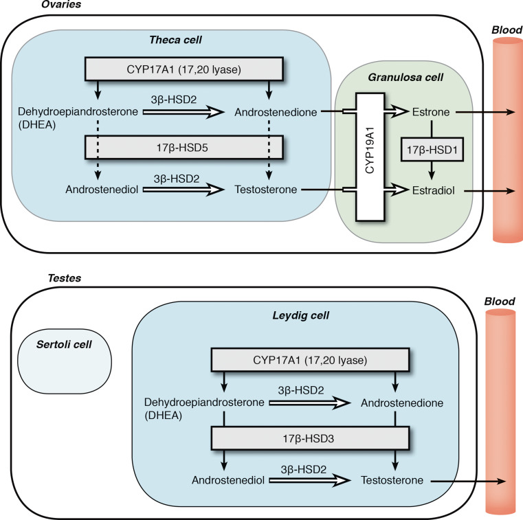
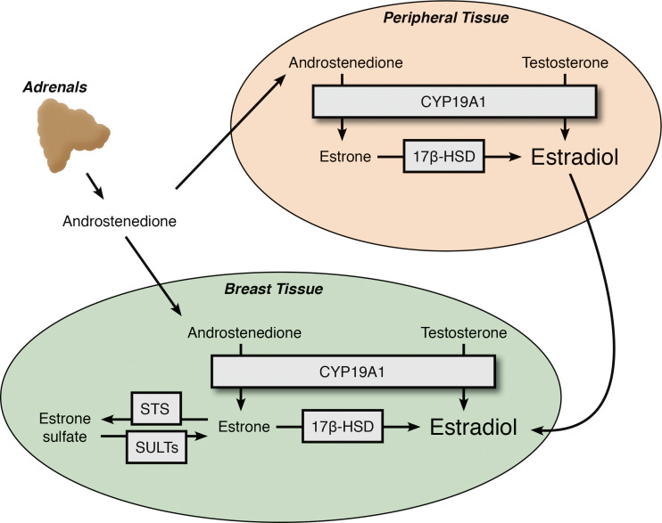
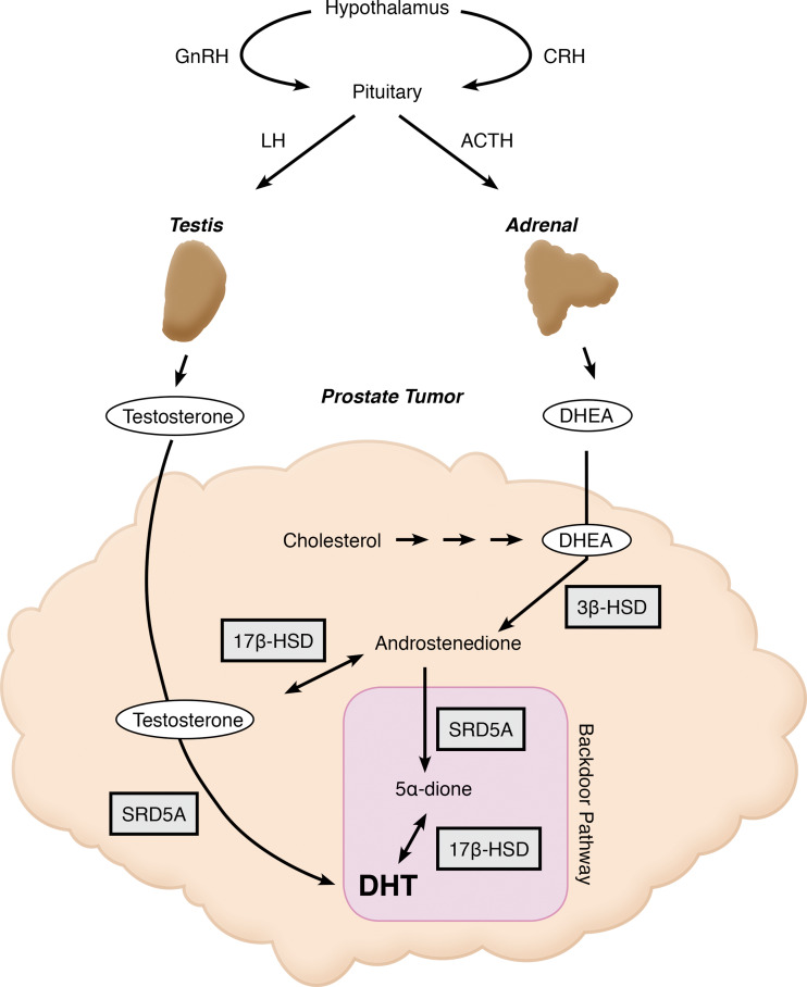
Breast and prostate cancers are malignancies in which steroid hormones drive cellular proliferation. Over the past century, this understanding has led to successful treatment strategies aimed to inhibit hormone-mediated tumor growth. Nonetheless, disease relapse and progression still pose significant clinical problems, with recurrent and metastatic tumors often exhibiting resistance to current drug therapies. The central role of androgens and estrogens in prostate and breast cancer etiology explains not only why endocrine therapies are often initially successful but also why many tumors ultimately become resistant. It is hypothesized that reducing the concentration of active hormones in the systemic circulation may be insufficient to block cancer progression, as this action selects for tumor cells that can generate active steroids from circulating precursors. This review aims to highlight the currently known differences of steroid biosynthesis in normal physiology versus hormone-dependent cancers, modern approaches to the assessment and targeting of these pathways, and priorities for future research.


Figures

Fig. 1
Fig. 1
Adrenal steroidogenesis. This schematic illustrates the biosynthesis pathways of mineralocorticoids, glucocorticoids, and sex steroids in the adrenal cortex by highlighting the predominant substrates and products within each zone. The three zones, zona glomerulosa (ZG), zona fasiculata (ZF), and zona reticularis (ZR), are labeled and designated with different background colors. Boxes denote steroidogenic enzymes, and arrows represent directionality of the enzymatic reactions. The pathway begins in the upper left hand corner with the conversion of cholesterol to pregnenolone. Multistep conversions are indicated with multiple arrows when the enzymes are not specified
Fig. 2
Fig. 2
Gonadal steroidogenesis. This figure depicts the enzymes expressed in the cells that comprise the gonads of females and males. a Ovarian theca cells express CYP17A1 to produce androstenedione and a small amount of testosterone, and these androgens are further aromatized into estrogens in ovarian granulosa cells before entering the circulation. b The testicular Leydig cells are the major steroidogenic cells in the male gonads, and these cells express CYP17A1 to convert androgen precursors into testosterone. Note the different 17βHSD isoenzymes present in the ovary and testis, which afford the major products E2 and testosterone, respectively
Fig. 3
Fig. 3
Sources of estrogen in postmenopausal women. Extragonadal estrogen secretion via intracrine and paracrine pathways significantly contributes to breast cancer progression. Breast tissue, in addition to other peripheral tissues, expresses CYP19A1 that mediates the conversion of circulating androgen precursors originating primarily in the adrenal, into E2. Following menopause, E2 remains a potent growth stimulus to the roughly 70 % of breast cancer cells expressing ER. AIs work by blocking the local conversion of androgens into estrogens in these extragonadal tissues and significantly reducing circulating E2 levels to prevent E2-induced tumor growth. E1S is an additional source of E2, as the local expression of estrone sulfatase (STS) is able to convert E1S back to the E2 precursor, E1
Fig. 4
Fig. 4
Pathways of androgen synthesis in CRPC. Shown in this figure are the pathways contributing to androgen synthesis that occur in the testes, adrenals, and prostate tumor itself in men with CRPC. Expression of CYP17A1 in all three tissues produces androgens, and further metabolism of these androgenic substrates results in the synthesis of the potent androgen receptor agonists, testosterone and DHT. Highlighted in purple is the backdoor pathway to DHT synthesis that bypasses testosterone as an intermediate. Independent studies have shown evidence to support this particular pathway being intact in CRPC

References

    1. Miller WL, Auchus RJ. The molecular biology, biochemistry, and physiology of human steroidogenesis and its disorders. Endocr Rev. 2011;32:81–151. doi: 10.1210/er.2010-0013. - DOI - PMC - PubMed
    1. American Cancer Society. Cancer facts & figures, 2015. Atlanta
    1. Vale W, Spiess J, Rivier C, Rivier J. Characterization of a 41-residue ovine hypothalamic peptide that stimulates secretion of corticotropin and beta-endorphin. Science. 1981;213:1394–1397. doi: 10.1126/science.6267699. - DOI - PubMed
    1. Rivier C, Vale W. Modulation of stress-induced ACTH release by corticotropin-releasing factor, catecholamines and vasopressin. Nature. 1983;305:325–327. doi: 10.1038/305325a0. - DOI - PubMed
    1. Catalano RD, Stuve L, Ramachandran J. Characterization of corticotropin receptors in human adrenocortical cells. J Clin Endocrinol Metab. 1986;62:300–304. doi: 10.1210/jcem-62-2-300. - DOI - PubMed

Publication types

MeSH terms