

Hedgehog regulates yes-associated protein 1 in regenerating mouse liver
- PMID: 26970079
- PMCID: PMC4917408
- DOI: 10.1002/hep.28542
Hedgehog regulates yes-associated protein 1 in regenerating mouse liver
Abstract
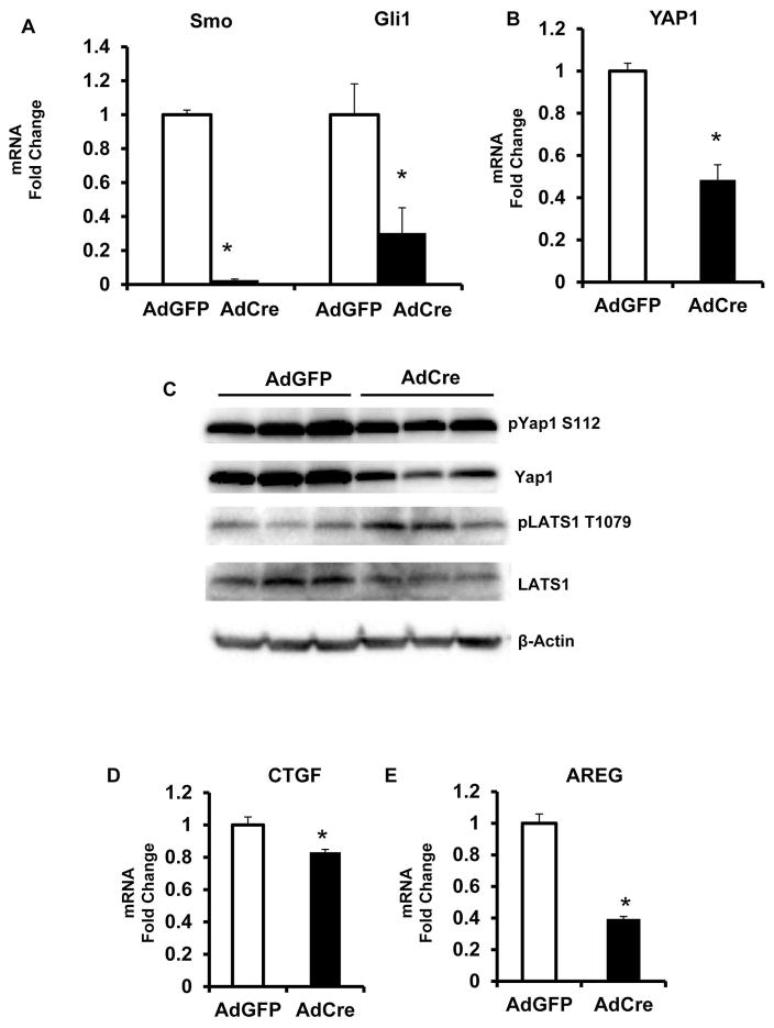
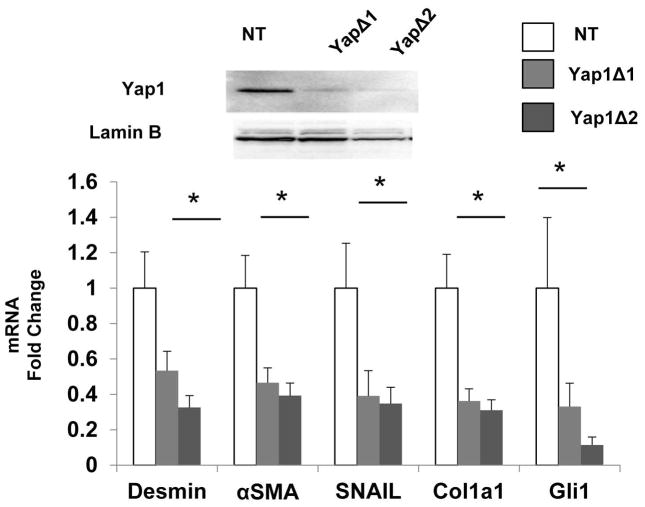
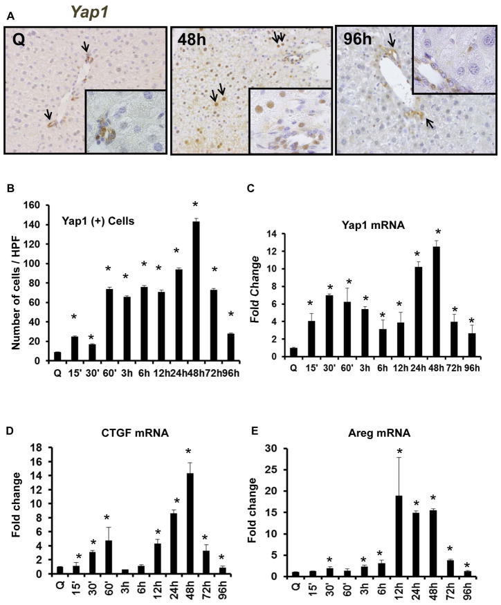
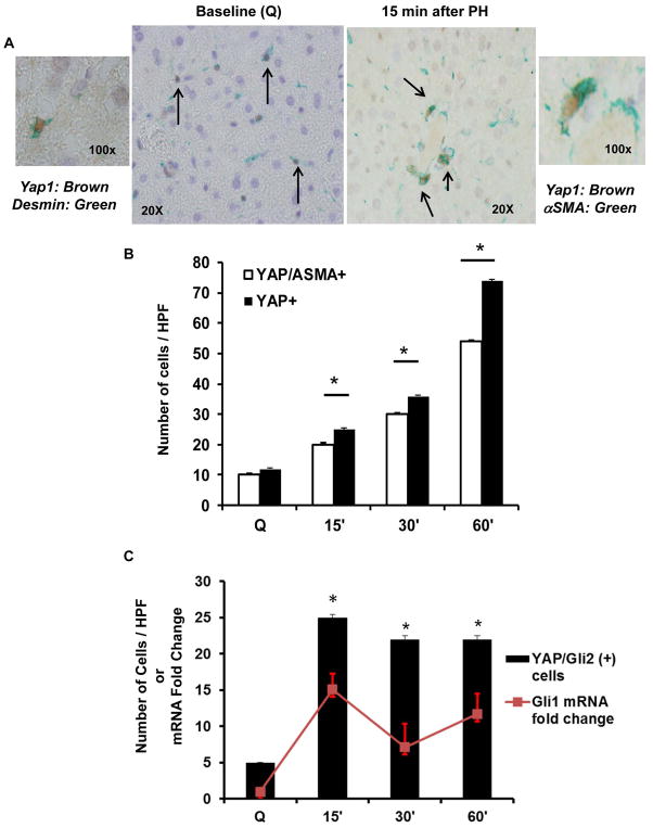
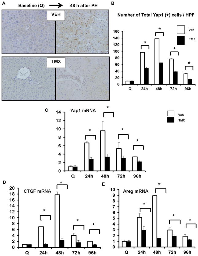
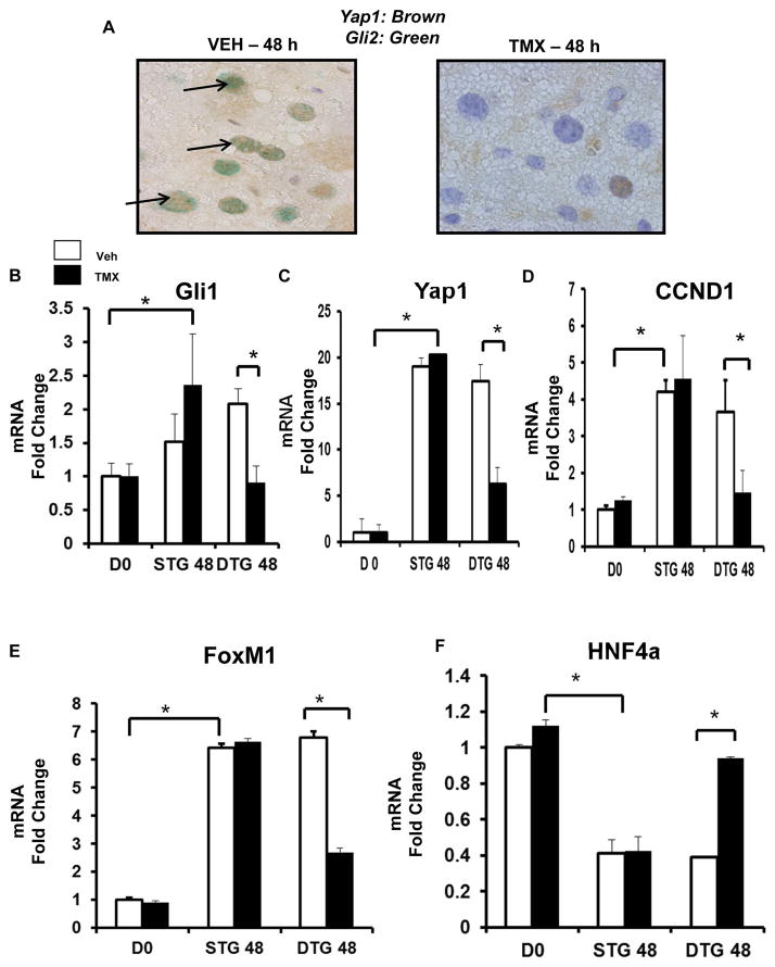
Adult liver regeneration requires induction and suppression of proliferative activity in multiple types of liver cells. The mechanisms that orchestrate the global changes in gene expression that are required for proliferative activity to change within individual liver cells, and that coordinate proliferative activity among different types of liver cells, are not well understood. Morphogenic signaling pathways that are active during fetal development, including Hedgehog and Hippo/Yes-associated protein 1 (Yap1), regulate liver regeneration in adulthood. Cirrhosis and liver cancer result when these pathways become dysregulated, but relatively little is known about the mechanisms that coordinate and control morphogenic signaling during effective liver regeneration. We evaluated the hypothesis that the Hedgehog pathway controls Yap1 activation during liver regeneration by studying intact mice and cultured liver cells. In cultured hepatic stellate cells (HSCs), disrupting Hedgehog signaling blocked activation of Yap1, and knocking down Yap1 inhibited induction of both Yap1- and Hedgehog-regulated genes that enable HSC to become myofibroblasts (MFs). In mice, disrupting Hedgehog signaling in MFs inhibited liver regeneration after partial hepactectomy (PH). Reduced proliferative activity in the liver epithelial compartment resulted from loss of stroma-derived paracrine signals that activate Yap1 and the Hedgehog pathway in hepatocytes. This prevented hepatocytes from up-regulating Yap1- and Hedgehog-regulated transcription factors that normally promote their proliferation.
Conclusions: Morphogenic signaling in HSCs is necessary to reprogram hepatocytes to regenerate the liver epithelial compartment post-PH. This discovery identifies novel molecules that might be targeted to correct defective repair during cirrhosis and liver cancer. (Hepatology 2016;64:232-244).
© 2016 by the American Association for the Study of Liver Diseases.
Conflict of interest statement
All authors declare no conflict of interest in relation to this study
None
Figures
Similar articles
-
Hedgehog-YAP Signaling Pathway Regulates Glutaminolysis to Control Activation of Hepatic Stellate Cells.Gastroenterology. 2018 Apr;154(5):1465-1479.e13. doi: 10.1053/j.gastro.2017.12.022. Epub 2018 Jan 3. Gastroenterology. 2018. PMID: 29305935 Free PMC article.
-
Liver regeneration requires Yap1-TGFβ-dependent epithelial-mesenchymal transition in hepatocytes.J Hepatol. 2018 Aug;69(2):359-367. doi: 10.1016/j.jhep.2018.05.008. Epub 2018 May 23. J Hepatol. 2018. PMID: 29758331 Free PMC article.
-
Cross-talk between Notch and Hedgehog regulates hepatic stellate cell fate in mice.Hepatology. 2013 Nov;58(5):1801-13. doi: 10.1002/hep.26511. Epub 2013 Sep 30. Hepatology. 2013. PMID: 23703657 Free PMC article.
-
Hedgehog signaling pathway as key player in liver fibrosis: new insights and perspectives.Expert Opin Ther Targets. 2014 Sep;18(9):1011-21. doi: 10.1517/14728222.2014.927443. Epub 2014 Jun 17. Expert Opin Ther Targets. 2014. PMID: 24935558 Review.
-
The role of Hedgehog signaling in fibrogenic liver repair.Int J Biochem Cell Biol. 2011 Feb;43(2):238-44. doi: 10.1016/j.biocel.2010.10.015. Epub 2010 Nov 5. Int J Biochem Cell Biol. 2011. PMID: 21056686 Free PMC article. Review.
Cited by
-
MicroRNA-378 is involved in hedgehog-driven epithelial-to-mesenchymal transition in hepatocytes of regenerating liver.Cell Death Dis. 2018 Jun 18;9(7):721. doi: 10.1038/s41419-018-0762-z. Cell Death Dis. 2018. PMID: 29915286 Free PMC article.
-
Recent advances in the role of Yes-associated protein in dermatosis.Skin Res Technol. 2023 Mar;29(3):e13285. doi: 10.1111/srt.13285. Skin Res Technol. 2023. PMID: 36973973 Free PMC article. Review.
-
Extra- and Intra-Cellular Mechanisms of Hepatic Stellate Cell Activation.Biomedicines. 2021 Aug 14;9(8):1014. doi: 10.3390/biomedicines9081014. Biomedicines. 2021. PMID: 34440218 Free PMC article. Review.
-
Maladaptive regeneration - the reawakening of developmental pathways in NASH and fibrosis.Nat Rev Gastroenterol Hepatol. 2021 Feb;18(2):131-142. doi: 10.1038/s41575-020-00365-6. Epub 2020 Oct 13. Nat Rev Gastroenterol Hepatol. 2021. PMID: 33051603 Free PMC article. Review.
-
Aging reduces liver resiliency by dysregulating Hedgehog signaling.Aging Cell. 2022 Feb;21(2):e13530. doi: 10.1111/acel.13530. Epub 2022 Jan 4. Aging Cell. 2022. PMID: 34984806 Free PMC article.
References
-
- Friedman SL. Evolving challenges in hepatic fibrosis. Nat Rev Gastroenterol Hepatol. 2010 Aug;7(8):425–36. - PubMed
MeSH terms
Substances
Grants and funding
LinkOut - more resources
Full Text Sources
Other Literature Sources
Research Materials
