Review
. 2016 Jun;228(6):889-909.
doi: 10.1111/joa.12449. Epub 2016 Mar 11.

Diffusible iodine-based contrast-enhanced computed tomography (diceCT): an emerging tool for rapid, high-resolution, 3-D imaging of metazoan soft tissues

Affiliations
Review

Diffusible iodine-based contrast-enhanced computed tomography (diceCT): an emerging tool for rapid, high-resolution, 3-D imaging of metazoan soft tissues

Paul M Gignac et al. J Anat. 2016 Jun.

Abstract

Morphologists have historically had to rely on destructive procedures to visualize the three-dimensional (3-D) anatomy of animals. More recently, however, non-destructive techniques have come to the forefront. These include X-ray computed tomography (CT), which has been used most commonly to examine the mineralized, hard-tissue anatomy of living and fossil metazoans. One relatively new and potentially transformative aspect of current CT-based research is the use of chemical agents to render visible, and differentiate between, soft-tissue structures in X-ray images. Specifically, iodine has emerged as one of the most widely used of these contrast agents among animal morphologists due to its ease of handling, cost effectiveness, and differential affinities for major types of soft tissues. The rapid adoption of iodine-based contrast agents has resulted in a proliferation of distinct specimen preparations and scanning parameter choices, as well as an increasing variety of imaging hardware and software preferences. Here we provide a critical review of the recent contributions to iodine-based, contrast-enhanced CT research to enable researchers just beginning to employ contrast enhancement to make sense of this complex new landscape of methodologies. We provide a detailed summary of recent case studies, assess factors that govern success at each step of the specimen storage, preparation, and imaging processes, and make recommendations for standardizing both techniques and reporting practices. Finally, we discuss potential cutting-edge applications of diffusible iodine-based contrast-enhanced computed tomography (diceCT) and the issues that must still be overcome to facilitate the broader adoption of diceCT going forward.

Keywords: Lugol's iodine; X-ray micro-CT scanning; alcoholic iodine; destaining; radiographic contrast agents; three-dimensional imaging.


Figures

Figure 1
Figure 1
A phylogenetically and morphologically diverse sample of tetrapods imaged using diffusible iodine‐based contrast‐enhanced computed tomography (diceCT), demonstrating the diversity of soft tissue types (e.g. muscles, glands, nerves, epithelia, fat) that can be visualized using this technique. (A) Sagittal slice through a crocodylian hatchling [Alligator mississippiensis (American alligator)], showing that Lugol's iodine solution readily penetrates even the heavily keratinized integument of reptiles, allowing for the clear visualization of internal organs such as the brain, heart, and liver. (B) Frontal slice through the head of an adult amphibian [Rana sylvatica (wood frog)], illustrating the detailed anatomical relationships among small, intricate structures of the auditory and ocular systems. (C) Sagittal and transverse biplanar cutaway view of a 3‐D volume rendering of a mammal [Mus musculus (house mouse)] embryo (15.5 days), showing the clarity with which minute developing structures can be imaged using diceCT. (D) Sagittal slice through a hatchling bird [Tyto alba (barn owl)], showing the completeness of whole‐body staining for post‐embryonic specimens. (E) Sagittal cutaway view of a 3‐D volume rendering of the head of an adult snake [Vipera berus (European adder)], showing digital reconstructions of the eye (blue), venom gland (yellow), ectopterygoid bone (white), and jaw adductor musculature (green). Specimens not to scale. Specimen preparation, staining, and scanning parameters can be found in Tables 1 and S1. Specimen images contributed by A.C.M., C.M.E., J.M., K.M., L.M.W., N.J.K., P.M.G., and R.M.H.
Figure 2
Figure 2
DiceCT imaging of American alligator (Alligator mississippiensis) hind limbs. (A) A 2‐D section through the acetabulum of a juvenile specimen (left), which was rendered into a single volume based on grayscale values (middle) and used as the basis for individually reconstructing limb muscles and bony elements in three dimensions (right). (B) A volumetric representation of a juvenile hind limb, sectioned through the proximal femoral metaphysis, demonstrating muscle bellies from ventral (top) and dorsal (bottom) views. (C) A close‐up view of the hip joint in an adult specimen, sectioned into an oblique anterolateral view and demonstrating acetabular soft tissues and oblique cartilages. acl, acetabular labrum; fc, fibrocartilage; fm, femur; hc, hyaline cartilage; il, ilium; mADD, m. adductor femoralis (parts 1 and 2); mAMB, m. ambiens; mCFB, m. caudofemoralis brevis; mCFL, m. caudofemoralis longus; mFT, m. femorotibialis; mFTE, m. flexor tibialis externus; mFTI, m. flexor tibialis internus; mIFB, m. iliofibularis; mIFM, m. iliofemoralis; mIT, m. iliotibialis; mPIFE, m. puboischiofemoralis externus (parts 1–3); pb, pubis; rac, rostral acetabular cartilage; sr, sacral rib. Specimen preparation, staining, and scanning parameters can be found in Tables 1 and S1. Specimen images contributed by C.M.H. and H.P.T.
Figure 3
Figure 3
DiceCT Do's: considerations and recommendations for successful specimen preparation.
Figure 4
Figure 4
Frontal diceCT slices through the heads of (A) a platypus (Ornithorhynchus anatinus; anterior to left) and (B) a western diamondback rattlesnake (Crotalus atrox; anterior to right). To ensure that both specimens are comparable, the grayscale ranges for A and B have been shifted so that the white values for the lenses (the whitest homologous structures of both specimens) are approximately equal. The platypus specimen was stored in 70% ethanol for more than 70 years and illustrates how the solubility of lipids in alcohol can reduce the potential for differentiation between different types of soft tissues in diceCT imaging of alcoholic specimens. In contrast, the rattlesnake was freshly fixed in 10% neutral buffered formalin, then stained and imaged shortly thereafter. Specimens are not to scale. Specimen preparation, staining, and scanning parameters can be found in Tables 1 and S1. Specimen images contributed N.J.K. and P.M.G..
Figure 5
Figure 5
DiceCT Don'ts: situations to avoid for successful specimen preparation.
Figure 6
Figure 6
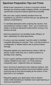
Specimen Preparation Tips and Tricks: a compilation of methodological shortcuts and timesaving measures from the authors’ collective experience, designed to help facilitate successful preparation and staining of diceCT specimens.
Figure 7
Figure 7
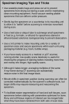
Specimen Imaging Tips and Tricks: a compilation of methodological shortcuts and timesaving measures from the authors’ collective experience, designed to help facilitate successful CT scanning of diceCT specimens.
Figure 8
Figure 8
Alternative perfusion‐based (A) and diffusion‐based (B, C [right], D) methods for enhancing soft‐tissue visualizations compared with diceCT imaging techniques (C [left], D). (A) A 3‐D volume rendering of the cranial vasculature of an African gray parrot (Psittacus erithacus; anterior is left) that was perfused with BriteVu. (B) Sagittal slice through the head of a domestic cat (Felis catus; anterior is left) stained with phosphomolybdic acid (PMA), demonstrating nasal and laryngeal cartilages and lingual musculature. (C) DiceCT image (left) of the baculum of a common pipistrelle bat (Pipistrellus pipistrellus) compared with a histological section (right) of the same specimen (anterior is top; modified from Herdina et al. 2015a,b). (D) Posterolateral view of a 3‐D volume rendering of the anterior portion of the head of a common pheasant (Phasianus colchicus; anterior toward the left) prepared using diceCT imaging (diffusible) for comparison to (A) and (B). Vasculature is particularly well visualized using injection techniques (A), whereas other tissues and spaces within the body cannot be readily imaged using this technique. Phosphomolybdic acid (B) stains muscle and hyaline cartilage, allowing for clear resolution of fiber attachment locations; while it also stains neural tissue, poor penetration through the cranium leaves the brain unstained. Histological preparations (C) are capable of targeting tissues with great specificity but are time‐consuming and difficult to translate into 3‐D. Further documentation for a wide range of alternative contrast agents, including the categories of histological tissues that can be readily visualized for each, can be found in Table S3. Preparation, staining, and scanning parameters for diceCT specimens (C, D) can be found in Tables 1 and S1. Specimen images contributed by A.N.H., C.P.O., J.A.C., M.S.E., and Z.L.

References

    1. Akkari N, Enghoff H, Metscher BD (2015) A new dimension in documenting new species: high‐detail imaging for myriapod taxonomy and first 3D cybertype of a new millipede species (Diplopoda, Julida, Julidae). PLoS ONE 10, e0135243. - PMC - PubMed
    1. Aslanidi OV, Nikolaidou T, Zhao J, et al. (2013) Application of micro‐computed tomography with iodine staining to cardiac imaging, segmentation, and computation model development. IEEE Trans Med Imaging 32, 8–17. - PMC - PubMed
    1. Balanoff AM, Bever GS, Colbert MW et al. (2015) Best practices for digitally constructing endocranial casts: examples from birds and their dinosaurian relatives. J Anat doi: 10.1111/joa.12378. - DOI - PMC - PubMed
    1. Baverstock H, Jeffery NS, Cobb SN (2013) The morphology of the mouse masticatory musculature. J Anat 223, 46–60. - PMC - PubMed
    1. Buytaert J, De Greef D, Aerts P, et al. (2014) Volume shrinkage of bone, brain and muscle tissue in sample preparation for micro‐CT and light sheet florescence microscopy (LSFM). Microsc Microanal 20, 1208–1217. - PubMed

Publication types