. 2017 Jan;37(1):36-48.
doi: 10.1177/0333102416637832. Epub 2016 Jul 11.

Sex-, stress-, and sympathetic post-ganglionic-dependent changes in identity and proportions of immune cells in the dura

Affiliations

Sex-, stress-, and sympathetic post-ganglionic-dependent changes in identity and proportions of immune cells in the dura

Lisa A McIlvried et al. Cephalalgia. 2017 Jan.

Abstract

Aim of investigation Due to compelling evidence in support of links between sex, stress, sympathetic post-ganglionic innervation, dural immune cells, and migraine, our aim was to characterize the impacts of these factors on the type and proportion of immune cells in the dura. Methods Dural immune cells were obtained from naïve or stressed adult male and female Sprague Dawley rats for flow cytometry. Rats with surgical denervation of sympathetic post-ganglionic neurons of the dura were also studied. Results Immune cells comprise ∼17% of all cells in the dura. These included: macrophages/granulocytes ("Macs"; 63.2% of immune cells), dendritic cells (0.88%), T-cells (4.51%), natural killer T-cells (0.51%), natural killer cells (3.08%), and B-cells (20.0%). There were significantly more Macs and fewer B- and natural killer T-cells in the dura of females compared with males. Macs and dendritic cells were significantly increased by stress in males, but not females. In contrast, T-cells were significantly increased in females with a 24-hour delay following stress. Lastly, Macs, dendritic cells, and T-cells were significantly higher in sympathectomized-naïve males, but not females. Conclusions It may not only be possible, but necessary to use different strategies for the most effective treatment of migraine in men and women.

Keywords: Headache; autonomic; inflammation; meninges.


Figures

Figure 1
Figure 1
Experimental design. (a) Intact and SCGx male and female rats were either considered “naïve” or stressed with the “CVS” paradigm. Tissue was collected from CVS rats immediately after the last day of stress (“CVS+0 h”) as a measure of the response to chronic stress or following a 24-hour delay after stress (“CVS+24 h”) as a measure of the response to stress-relaxation. (b) To test the impact of sympathetic post-ganglionic neuron innervation, prior to stress, superior cervical ganglia were surgically removed bilaterally (SCGx) in the groups as indicated. On the left, a representative whole-mount dura from an adult rat 7 days after a unilateral SCGx is shown, probed with anti-CGRP (green) or anti-TH (red) antibodies. Note the complete loss of TH-like immunoreactivity (a marker for sympathetic fibers) ipsilateral to the SCGx, but no change in CGRP-like immunoreactivity (a marker for peptidergic afferents). (c) CVS is a mild stress paradigm. There was a significant (p<0.01) main effect of stress on plasma levels of CORT in rats. CORT was significantly (p<0.01) higher in rats exposed to CVS (naïve versus CVS+0 h), and this remained significantly (p<0.01) elevated with a delay after stress (naïve versus CVS+24 h). There was no significant (p>0.05) main effect of sex or interaction with stress. Note that the CVS paradigm produced a significantly (p<0.001) smaller increase in CORT compared with the potent, acute restraint stress paradigm. (d) In addition, there was a significant (p<0.001) main effect of stress on daily weight gain in rats. Rats gained significantly (p<0.05) less weight per day during CVS (naïve versus CVS+0 h), which recovered (p<0.001) during the relaxation phase after stress (CVS+0 h versus CVS+24 h). CGRP: calcitonin gene-related peptide; CORT: corticosterone; CVS: chronic variable stress; SCGx: surgical sympathectomy; TH: tyrosine hydroxylase.
Figure 2
Figure 2
Gating strategy used for the isolation of immune cell subtypes from the dura. The data shown are from a naïve male rat. On the day of the experiment, dural cells were dissociated and stained with antibodies specific to markers of immune cells and/or distinct immune cell subtypes. The gating strategy illustrated was used for all flow and fluorescence-activated cell sorting experiments. CD45+ immune cells were first selected, and then sorted into CD11b+ macrophages/granulocytes/mast cells (“Macs”) and CD11c+DCs. CD11b/c immune cells were further sorted into CD45R+ B-cells. CD11b/c and CD45R immune cells were lastly sorted into either CD161a+ NK cells, CD3+ T-cells, or CD161a+/CD3+ NKT cells. A small subset of immune cells was left unstained by any of the immune subtype antibodies that were utilized in this strategy (“unidentified”). Viability was uniformly 80–85%, as determined by propidium iodide and trypan blue staining. This resulted in an average of 1.56×10⁶±0.18×10⁶ live cells recovered per dura. Spleen cells were used for compensation controls (51,52). Images are from FlowJo software, contour plots were used with 5% levels and outliers displayed. DC: dendritic cell; NK: natural killer; FSC-A: forward scatter.
Figure 3
Figure 3
Proportion of immune cell subtypes in the naïve male dura as determined with flow cytometry using the gating strategy shown in Figure 2. Of the live cells recovered from the dura, an average of 16.9±0.90% were CD45+, and thus determined to be immune cells. The pie chart and legend show the relative proportions of the six immune cell subtypes identified in the dura. The insets show fluorescence-activated cell sorted, cyto-centrifuged preparations of immune cell subtypes, subsequently stained with Diff-Quik. Macs consisted of 89.1% macrophages/monocytes (M), 5.1% neutrophils (N), 3.6% mast cells (MC; identified with toluidine blue stain), and 2.2% lymphocytes. The NK cell population consisted of 51.3% large lymphocytes (>10 μm) and 48.7% small lymphocytes (≤10 μm). The T-cell population consisted of 79.7% small lymphocytes and 20.3% large lymphocytes. Lastly, the B-cell population consisted of 93.0% small lymphocytes, 5.1% lymphocytes, and 1.9% macrophages/monocytes. Scale bars: 10 μm. DC: dendritic cell; Macs: macrophages/granulocytes; NK: natural killer.
Figure 4
Figure 4
Sex-, stress-, and SPGN-dependent changes in the percentage of CD45+ immune cells in the dura (relative to the total number of live cells recovered). Data are presented as percentages of naïve males in order to facilitate comparisons between groups. (a) There was a significant interaction between sex and stress regarding the proportion of live immune cells in the dura (p<0.05) (“Intact” groups). In addition, there was a significant (p<0.05) interaction between sex and SPGN innervation in naïve animals regarding the proportions of immune cells in the dura (“Intact, naïve” and “SCGx, naïve” groups). Lastly, there was an interaction between sex, stress, and SPGN innervation (p<0.05) regarding the proportion of immune cells in the dura. (b) Similar to male SCGx groups, male sham-operated rats also showed the loss of a stress-induced increase in immune cells. H1=a priori hypothesis 1: sex difference in “Intact, naïve” groups, analyzed with a t-test; H2=a priori hypothesis 2: sex×stress comparison in “Intact” groups, analyzed with a two-way analysis of variance (ANOVA); H3=a priori hypothesis 3: sex×SCGx in “Naïve” groups (i.e. “Intact” versus “SCGx” naïve), analyzed with a two-way ANOVA; H4=a priori hypothesis 4: sex×stress SCGx interaction between all groups, analyzed with a three-way ANOVA. *p<0.05. CVS: chronic variable stress; SCGx: surgical sympathectomy; SPGN: sympathetic post-ganglionic neuron.
Figure 5
Figure 5
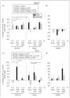
Sex-, stress-, and SPGN-dependent changes in the percentage of myeloid-derived immune cells dissociated from the dura (relative to the total number of live cells from each dura). Data are presented as percentages of naïve males for comparison. (a) The proportion of Macs was significantly (p<0.05) higher in the dura from intact naïve females than males. There was a significant (p<0.05) interaction between sex and stress regarding the proportion of Macs in the dura (“Intact” groups). There was also a significant (p<0.05) interaction between sex and SPGN innervation in naïve rats regarding the proportion of Macs in the dura (“Intact, naïve” and “SCGx, naïve” groups). Lastly, there was an interaction between sex, stress, and SPGN innervation (p<0.05). (b) Similar to male SCGx groups, male sham-operated rats also showed the loss of a stress-induced increase in Macs. (c) There was a significant (p<0.05) interaction between sex and stress on the proportion of dural DCs (“Intact” groups). In addition, there was a significant (p<0.05) interaction between sex, stress, and SCGx. (d) Interestingly, in sham rats, the stress-induced increase in DCs was only seen with a delay following stress. The groups and a priori hypotheses (H1–4) tested were the same as those in Figure 4. *p<0.05; **p<0.01. CVS: chronic variable stress; DC: dendritic cell; Macs: macrophages/granulocytes; SCGx: surgical sympathectomy; SPGN: sympathetic post-ganglionic neuron.
Figure 6
Figure 6
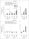
Sex-, stress-, and SPGN-dependent changes in the percentage of lymphoid-derived immune cells dissociated from the dura (relative to the total number of live cells from each dura). Data are presented as percentages of naïve males for comparison. (a) There was a significant (p<0.05) interaction between sex and stress regarding the relative proportions of T-cells (“Intact” groups). In addition, there was a significant main effect (p<0.05) of SPGN innervation in naïve rats. Lastly, the sex and stress interaction and SPGN main effect persisted with all groups folded into the analysis. (b) Comparable stress-induced changes in T-cells were observed in sham surgery groups and in the SCGx groups. (c) There was a main effect of sex (p<0.05) on the proportion of NKT cells. (d) Sham-operated groups showed a stress effect in males, which may have been masked by SCGx. (e) There was a significant (p<0.05) sex and stress interaction when all groups were folded into the analysis of NK cells. (f) Sham surgery did not appear to affect NK cells. (g) The proportion of B-cells was significantly (p<0.001) lower in the dura from intact naïve females than males, and this sex difference persisted as a main effect (p<0.05) after folding stress groups into the analysis (“Intact” groups). In addition, when all groups were folded in, there was a significant (p<0.05) main effect of SPGN innervation. (h) Sham surgery did not appear to affect B-cells. The groups and a priori hypotheses (H1–4) tested were the same as those in Figure 4. *p<0.05; **p<0.01. CVS: chronic variable stress; DC: dendritic cell; NK: natural killer; SCGx: surgical sympathectomy; SPGN: sympathetic post-ganglionic neuron.
Figure 6
Figure 6
Sex-, stress-, and SPGN-dependent changes in the percentage of lymphoid-derived immune cells dissociated from the dura (relative to the total number of live cells from each dura). Data are presented as percentages of naïve males for comparison. (a) There was a significant (p<0.05) interaction between sex and stress regarding the relative proportions of T-cells (“Intact” groups). In addition, there was a significant main effect (p<0.05) of SPGN innervation in naïve rats. Lastly, the sex and stress interaction and SPGN main effect persisted with all groups folded into the analysis. (b) Comparable stress-induced changes in T-cells were observed in sham surgery groups and in the SCGx groups. (c) There was a main effect of sex (p<0.05) on the proportion of NKT cells. (d) Sham-operated groups showed a stress effect in males, which may have been masked by SCGx. (e) There was a significant (p<0.05) sex and stress interaction when all groups were folded into the analysis of NK cells. (f) Sham surgery did not appear to affect NK cells. (g) The proportion of B-cells was significantly (p<0.001) lower in the dura from intact naïve females than males, and this sex difference persisted as a main effect (p<0.05) after folding stress groups into the analysis (“Intact” groups). In addition, when all groups were folded in, there was a significant (p<0.05) main effect of SPGN innervation. (h) Sham surgery did not appear to affect B-cells. The groups and a priori hypotheses (H1–4) tested were the same as those in Figure 4. *p<0.05; **p<0.01. CVS: chronic variable stress; DC: dendritic cell; NK: natural killer; SCGx: surgical sympathectomy; SPGN: sympathetic post-ganglionic neuron.
Figure 7
Figure 7
Sex-, stress-, and SPGN-dependent changes in the percentage of unidentified immune cells dissociated from the dura (relative to the total number of live cells from each dura). Data are presented as percentages of naïve males for comparison. (a) With all groups folded into the analysis, there was a main effect of sex (p<0.05) on the proportion of “unidentified” immune cells. (b) Sham surgery did not appear to affect “unidentified” cells. The groups and a priori hypothesis (H4) tested were the same as those in Figure 4. CVS: chronic variable stress; SCGx: surgical sympathectomy; SPGN: sympathetic post-ganglionic neuron.

References

    1. Levy D. Endogenous mechanisms underlying the activation and sensitization of meningeal nociceptors: the role of immuno-vascular interactions and cortical spreading depression. Curr Pain Headache Rep. 2012;16:270–277. - PubMed
    1. Olesen J, Burstein R, Ashina M, et al. Origin of pain in migraine: evidence for peripheral sensitisation. Lancet Neurol. 2009;8:679–690. - PubMed
    1. Waeber C, Moskowitz MA. Migraine as an inflammatory disorder. Neurology. 2005;64(10 Suppl. 2):S9–S15. - PubMed
    1. Levy D, Burstein R, Kainz V, et al. Mast cell degranulation activates a pain pathway underlying migraine headache. Pain. 2007;130:166–176. - PMC - PubMed
    1. Reuter U, Bolay H, Jansen-Olesen I, et al. Delayed inflammation in rat meninges: implications for migraine pathophysiology. Brain. 2001;124:2490–2502. - PubMed

LinkOut - more resources