Review
. 2016:116:115-34.
doi: 10.1016/bs.ctdb.2015.12.003. Epub 2016 Jan 23.

The Neural Crest Migrating into the Twenty-First Century

Affiliations
Review

The Neural Crest Migrating into the Twenty-First Century

Marianne E Bronner et al. Curr Top Dev Biol. 2016.

Abstract

From the initial discovery of the neural crest over 150 years ago to the seminal studies of Le Douarin and colleagues in the latter part of the twentieth century, understanding of the neural crest has moved from the descriptive to the experimental. Now, in the twenty-first century, neural crest research has migrated into the genomic age. Here, we reflect upon the major advances in neural crest biology and the open questions that will continue to make research on this incredible vertebrate cell type an important subject in developmental biology for the century to come.

Keywords: Craniofacial skeleton; Embryo; Neural crest; Peripheral nervous system; Vertebrates.


Figures

Figure 1
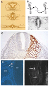
Figure 1. Neural crest cells delaminate from the neural tube and undergo extensive migration
(A) Immunohistochemistry with an antibody for neural crest marker Pax7 highlights neural crest cells delaminating from the neural tube of a stage HH9 chicken embryo. (B) Diagram representing a cross section of a neurulating chicken embryo. At the stage represented, the neural cells occupy the dorsal portion of the neural tube. (C) Neural crest cells eventually undergo epithelial to mesenchymal transition to delaminate from the neural tube and undergo extensive migration.
Figure 2
Figure 2. Classical experiments in neural crest research utilizing avian embryos
(A) Reproduction of the original drawings of Wilhelm His, including the first depiction of neural crest cells (His, 1898). The neural crest is identified with a “Z” for "Zwischenstrang” or cord between the neural and non-neural ectoderm. (B) Transplant experiments by Weston (1963), where neural tubes from radioactively labeled embryos were transplanted to unlabeled hosts. This allowed for the tracking of neural crest cells after delamination and identification of distinct migratory pathways. (C) The chick-quail chimeras of Nicole Le Douarin. Grafting of quail tissue in chicken embryos permitted extensive fate mapping of progenitor cells (Le Douarin, 2004). This approach was used to map migratory pathways and derivatives of the neural crest at distinct axial levels of the avian embryo (Le Douarin, 1982). (D) Clonal analysis of neural crest cells in the developing embryo. Labeling of single neural crest cells with vital dyes showed that many individual progenitor cells are multipotent and differentiate into diverse cell types in vivo (Fraser and Bronner-Fraser, 1991).
Figure 3
Figure 3. A gene regulatory network (GRN) controls neural crest formation
A complex genetic program comprised of numerous transcription factors underlies neural crest development. These factors are arranged in distinct hierarchical modules corresponding to the different stages of neural crest formation. First, in the neural plate border, the interplay of signaling systems such as Wnts, Bmps and Fgfs activates the neural plate border specifier genes, which include Gbx2, Msx1, Zic1, Tfap2 and Pax7. These genes drive the transition of the GRN to the neural crest specification module. Signaling systems such as Wnts also feed into the system by activating transcription factor Axud1, which interacts with PAX7 and MSX1 to activate expression of neural crest specifier genes (FoxD3, Ets1, Sox9, etc.) in the dorsal neural folds. The neural crest specifier genes endow the neural crest with its defining features and activate the process of epithelial to mesenchymal transition. In the migratory neural crest, there is activation of additional factors that will affect migration and drive differentiation in multiple cell types.

Similar articles

Cited by

References

    1. Abercrombie M, Heaysman JE. Observations on the social behaviour of cells in tissue culture: I. Speed of movement of chick heart fibroblasts in relation to their mutual contacts. Exp Cell Res. 1954;5:111–131. - PubMed
    1. Abitua PB, Wagner E, Navarrete IA, Levine M. Identification of a rudimentary neural crest in a non-vertebrate chordate. Nature. 2012;492:104–107. - PMC - PubMed
    1. Adameyko I, Lallemend F, Aquino JB, Pereira JA, Topilko P, Müller T, Fritz N, Beljajeva A, Mochii M, Liste I, Usoskin D, Suter U, Birchmeier C, Ernfors P. Schwann cell precursors from nerve innervation are a cellular origin of melanocytes in skin. Cell. 2009;139:366–379. - PubMed
    1. Baggiolini A, Varum S, Mateos JM, Bettosini D, Nessy J, Ziegler U, Dimou L, Clevers H, Furrer R, Sommer L. Genetic Lineage Tracing Demonstrates Multipotency of Premigratory and Migratory Neural Crest Cells in Vivo. Cell Stem Cell. 2015;6(3):314–322. - PubMed
    1. Baroffio A, Dupin E, Le Douarin NM. Clone-forming ability and differentiation potential of migratory neural crest cells. Proc Natl Acad Sci USA. 1988;85:5325–5329. - PMC - PubMed

LinkOut - more resources