Review
. 2016:116:315-30.
doi: 10.1016/bs.ctdb.2015.11.001. Epub 2016 Feb 27.

Connectomics, the Final Frontier

Affiliations
Review

Connectomics, the Final Frontier

Scott W Emmons. Curr Top Dev Biol. 2016.

Abstract

How the synaptic connections in the nervous system are genetically encoded and formed during development remains an unsolved problem. The known connectivity of the nervous system of the nematode C. elegans provides an opportunity to search for the genes involved. The circuits for male mating behavior form a complex neural network that would seem to require a large family of molecular cell labels for pre- and postsynaptic cell recognition. It is suggested that a combinatorial code of neural cell adhesion proteins specifying the network of connections may be discovered by comparing the expression patterns of candidate genes with the pattern of connections.

Keywords: C. elegans; Combinatorial code; Nervous system; Nervous system development; Neural cell adhesion protein; Neural network; Synapse; Synaptic connectivity.


Figures

Figure 1
Figure 1
The nerve bundles of the C. elegans nervous system, visualized by a reporter gene that expresses the fluorescent protein GFP in all of the neurons. (Image from Hang Ung, Jean-Louis Bessereau laboratory, France.)
Figure 2
Figure 2
Ganglia in the C. elegans male tail. CG(L), left cloacal ganglion; DNC, dorsal nerve cord; DRG, dorsorectal ganglion; LG(L), left lumbar ganglion; LG(R), right lumbar ganglion; PAG, preanal ganglion; VNC, ventral nerve cord; right cloacal ganglion not shown. Cell bodies are distributed in each of the six ganglia. Synapses can be found throughout, but are situated primarily within the PAG. Neurons outside this ganglion send processes through commissures into it. (Image from WormAtlas, http://www.wormatlas.org.)
Figure 3
Figure 3
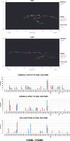
Homologous neurons have dissimilar branching structures in the preanal ganglion but similar sets of synaptic partners. Upper panels: the processes and synapses in the PAG of a pair of homologous neurons, the ray sensory neurons R2BL and R2BR. The cell bodies of these neurons are respectively in the left and right lumbar ganglia (Figure 2). Pink circles show sites of chemical synaptic output, magenta circles are chemical input, and blue circles are gap junctions. These maps are from Wormwiring.org. Lower panels: histograms comparing the synaptic relationships made by R2BL and R2BR. The neurons listed along the bottom are the 68 neurons (out of 170) with synaptic interaction of some kind with one or both of these two neurons Data for left/right homologs of R2BL and R2BR partners are combined; e.g. R1A represents the interaction with R1AL plus R1AR. The vertical axis gives the total morphological strength of interaction in number of EM serial sections. Note the differences in scales.
Figure 4
Figure 4
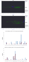
PVV and PDB are distinct neurons with similar patterns of synaptic input. Top panels show the overlapping branching structures of PVV and PDB in the preanal ganglion. PVV extends further anteriorly into the ventral cord, while PDB sends a process posteriorly around the end of the tail to the dorsal side. Both have output onto bodywall muscles. The blue boxes are the cell bodies. The horizontal axes show the serial section number of the transverse serial sections (A-P axis); the vertical axis shows position along the D-V axis; the maps are flattened along the L-R axis. The top histogram shows connectivity data for all the ray neurons; R1A(L/R), R2A(L/R), R3A(L/R) and R6B(L/R) make no connection to either PVV or PDB. The bottom histogram shows data for all the remaining neurons that make a chemical connection to either neuron. Data for left/right homologs and other equivalent neurons are combined.

References

    1. Brenner S. The genetics of Caenorhabditis elegans. Genetics. 1974;77:71–94. - PMC - PubMed
    1. Brenner S. In the Beginning Was the Worm …. Genetics. 2009;182:413–415. - PMC - PubMed
    1. Chalfie M, Sulston JE, White JG, Southgate E, Thomson JN, Brenner S. The neural circuit for touch sensitivity in Caenorhabditis elegans. The Journal of Neuroscience. 1985;5:956–964. - PMC - PubMed
    1. Chia PH, Li P, Shen K. Cellular and molecular mechanisms underlying presynapse formation. The Journal of Cell Biology. 2013;203:11–22. - PMC - PubMed
    1. Colon-Ramos DA. Current Topics in Developmental Biology. Academic Press; 2009. Chapter 2 Synapse Formation in Developing Neural Circuits; pp. 53–79. - PMC - PubMed

Publication types

Substances

LinkOut - more resources