. 2016 May:492:232-41.
doi: 10.1016/j.virol.2016.02.019. Epub 2016 Mar 21.

Mapping and modeling of a strain-specific epitope in the Norwalk virus capsid inner shell

Affiliations

Mapping and modeling of a strain-specific epitope in the Norwalk virus capsid inner shell

Gabriel I Parra et al. Virology. 2016 May.

Abstract

Noroviruses are diverse positive-strand RNA viruses associated with acute gastroenteritis. Cross-reactive epitopes have been mapped primarily to conserved sequences in the capsid VP1 Shell (S) domain, and strain-specific epitopes to the highly variable Protruding (P) domain. In this work, we investigated a strain-specific linear epitope defined by MAb NV10 that was raised against prototype (Genogroup I.1) strain Norwalk virus (NV). Using peptide scanning and mutagenesis, the epitope was mapped to amino acids 21-32 (LVPEVNASDPLA) of the NV S domain, and its specificity was verified by epitope transfer and reactivity with a recombinant MAb NV10 single-chain variable fragment (scFv). Comparative structural modeling of the NV10 strain-specific and the broadly cross-reactive TV20 epitopes identified two internal non-overlapping sites in the NV shell, corresponding to variable and conserved amino acid sequences among strains, respectively. The S domain, like the P domain, contains strain-specific epitopes that contribute to the antigenic diversity among the noroviruses.

Keywords: Caliciviruses; Epitope-tagging; Noroviruses.


Figures

Fig. 1.
Fig. 1.
Definition of five antigenic sites on the Norwalk virus (NV) VP1 by competitive binding assays with monoclonal antibodies (MAbs). Matrix summarizes cross-competition among P-domain NV MAbs. A black box indicates 450% blocking (positive competition) of unlabeled MAb against biotinylated MAbs, and a white box indicates no competition (o50% blocking). The VP1 binding domain for each MAb was determined by reactivity with a panel of GI/GII chimeric VLPs (5). Linear epitopes were screened by reactivity of the MAbs with the 58 kDa NV VP1 band in immunoblots and with overlapping peptide libraries representing the NV S and P domains. Antigenic site V (mapping to the S domain) was defined based on binding to GI/GII chimeric VLPs and the S peptide library. Carbohydrate-blocking activity for each MAb was measured by hemagglutination inhibition (HAI) assays. The lowest amount of MAb to achieve full inhibition of the hemagglutination of human red blood cells with NV VLPs is indicated. Positive HAI activity is indicated in red type. Experimental details are described in Section 3.
Fig. 2.
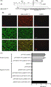
Fig. 2.
Localization of the NV10 epitope to the Shell domain of the VP1 protein. (A). Reactivity of MAb NV10 with a library of overlapping peptides corresponding to the Shell domain of NV. Positive peptides span residues 16–37 of the NV capsid protein. (B) Immunofluorescence staining of MAb NV10 with Huh-7 cells carrying the replicon of NV. Schematic diagram of the genome organization of the NV replicon construct showing the presence of the first 32 amino acid residues of the VP1 protein encoded within the beginning of the ORF2 (nucleotide 5358) to the BamH1 restriction enzyme site (nucleotide 5456) in ORF2 (Adapted from Chang et al. (2006)). Huh-7 cells without replicon are included as negative control. Experiments were performed as described in Section 3.
Fig. 3.
Fig. 3.
Reactivity of NV10 MAb with truncated NV VP1 proteins. (A) Design of deletions that were engineered into the NV VP1 protein encoded in the pCI eukaryotic expression vector. Amino acid numbers correspond to the NV VP1 protein. (B) Immunofluorescence staining reactivity of MAb NV10 with Vero cells transfected with pCI-based plasmid constructions containing the engineered deletions. Hyperimmune serum against NV VP1 [α-NV(VP1)] was used to verify expression of the constructs.
Fig. 4.
Fig. 4.
Fine mapping of NV10 MAb epitope. (A) Amino acid alignment (residues 11–32) from various norovirus VLPs tested with MAb NV10. Amino acid numbers correspond to the NV VP1 protein. The region (residues 21–32) shown to be critical for NV10 binding is indicated at the top. (B) Reactivity of NV10 with an alanine scanning peptide library that spans residues 11–32 of NV VP1. A peptide containing the wild-type residue was used as positive control. (C) Immunofluorescence staining results of NV10 with Vero cells transfected with pCI-based plasmid constructions containing mutations in the VP1 from NV. Hyperimmune serum against NV VP1 [α-NV(VP1)] was used to test expression of the constructs.
Fig. 5.
Fig. 5.
Molecular modeling of NV10 MAb and its cognate epitope. (A) Structure of the NV VP1 (PDB: 1IHM) protein showing the location of the putative epitope recognized by MAb NV10 (highlighted in green on the VP1 B monomer). The structural domains of the VP1 are indicated on the left-hand side. (B) Cross-section view of a reconstructed NV capsid showing the location of the epitope NV10 sequence and the highly cross-reactive epitope TV20 (highlighted in red) (Parra et al., 2013). Only one of the VP1 trimer monomeric subunits (B monomer) was used to illustrate the inner surface exposure of the epitopes. (C) Electrostatic potential on the surface of NV VP1 (Bottom View). Electrostatic potential was calculated and visualized using USCF Chimera (Pettersen et al., 2004). The NV10-epitope is indicated with a square. Electrostatic potential scale±10 kcal/mole. (D) Molecular model of the Fab region of MAb NV10. The left-hand side view shows the light and heavy chain in different colors, while the right-hand side view shows the electrostatic potential of the surface. The binding region is indicated with a square showing the electrostatic complementarities among the interacting surfaces. The binding region was obtained by modeling the NV10 Fab using Rosetta Antibody with default settings (Sivasubramanian et al., 2009).
Fig. 6.
Fig. 6.
Tagging heterologous proteins with NV10 epitope. (A) Schematic representation showing a recombinant feline calicivirus (FCV) clone (vR6-LC-VP111–32) in which the unique NV10 epitope was introduced into the leader of the capsid (LC) protein from FCV. The HA and NV10 epitopes introduced are underlined. (B) Immunofluorescence staining of CRFK cells infected with recombinant FCV carrying the NV10 epitope. Wild-type virus (vR6) and CRFK cells were used as controls. Hyperimmune serum raised against the LC protein (α-LC) verified that CRFK cells were infected with both FCV strains. (C) Results of the mammalian two-hybrid assay showing the specific interaction of the NV10 scFv and the NV VP1 protein. Plasmids are designated with the two-hybrid cloning vector listed first (pM or pVP16) followed by the name of the protein engineered for expression. Plasmids expressing the T antigen [pM(T)] and the p53 protein [pVP16(53)] were used as positive controls, as well as plasmids showing the VP1:VP1 interactions of NV and FCV. A plasmid expressing the CAT enzyme (pG5CAT), and empty plasmids (pVP16 and pM) served as negative controls. Interaction between the NV10 scFv and NV VP1 protein was evidenced by a positive signal in the pM(NV10scFv)+pV16(NV-VP1) two-hybrid experiment. NV10 scFv specificity was tested against FCV VP1 [pV16(FCV-VP1)].

Similar articles

Cited by

References

    1. Atmar RL, Bernstein DI, Harro CD, Al-Ibrahim MS, Chen WH, Ferreira J, Estes MK, Graham DY, Opekun AR, Richardson C, Mendelman PM, 2011. Norovirus vaccine against experimental human Norwalk virus illness. N. Engl. J. Med. 365, 2178–2187. - PMC - PubMed
    1. Abente EJ, Sosnovtsev SV, Sandoval-Jaime C, Parra GI, Bok K, Green KY, 2013. The feline calicivirus leader of the capsid protein is associated with cytopathic effect. J. Virol. 87, 3003–3017. - PMC - PubMed
    1. Atmar RL, Opekun AR, Gilger MA, Estes MK, Crawford SE, Neill FH, Graham DY, 2008. Norwalk virus shedding after experimental human infection. Emerg. Infect. Dis. 14, 1553–1557. - PMC - PubMed
    1. Bertolotti-Ciarlet A, Crawford SE, Hutson AM, Estes MK, 2003. The 3′ end of Norwalk virus mRNA contains determinants that regulate the expression and stability of the viral capsid protein VP1: a novel function for the VP2 protein. J. Virol. 77, 11603–11615. - PMC - PubMed
    1. Bok K, Parra GI, Mitra T, Abente E, Shaver CK, Boon D, Engle R, Yu C, Kapikian AZ, Sosnovtsev SV, Purcell RH, Green KY, 2011. Chimpanzees as an animal model for human norovirus infection and vaccine development. Proc. Natl. Acad. Sci. USA 108, 325–330. - PMC - PubMed

Publication types

MeSH terms