. 2016 Sep;138(3):825-838.
doi: 10.1016/j.jaci.2015.12.1330. Epub 2016 Mar 11.

Clonal expansion of CD4(+) cytotoxic T lymphocytes in patients with IgG4-related disease

Affiliations

Clonal expansion of CD4(+) cytotoxic T lymphocytes in patients with IgG4-related disease

Hamid Mattoo et al. J Allergy Clin Immunol. 2016 Sep.

Abstract

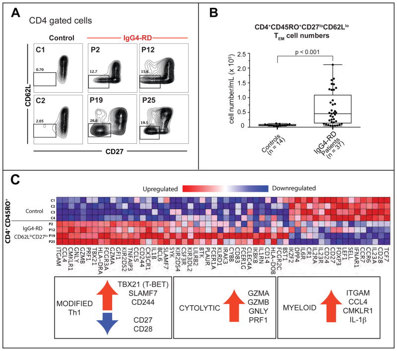
Background: IgG4-related disease (IgG4-RD) is a systemic condition of unknown cause characterized by highly fibrotic lesions with dense lymphoplasmacytic infiltrates. CD4(+) T cells constitute the major inflammatory cell population in IgG4-RD lesions.

Objective: We used an unbiased approach to characterize CD4(+) T-cell subsets in patients with IgG4-RD based on their clonal expansion and ability to infiltrate affected tissue sites.

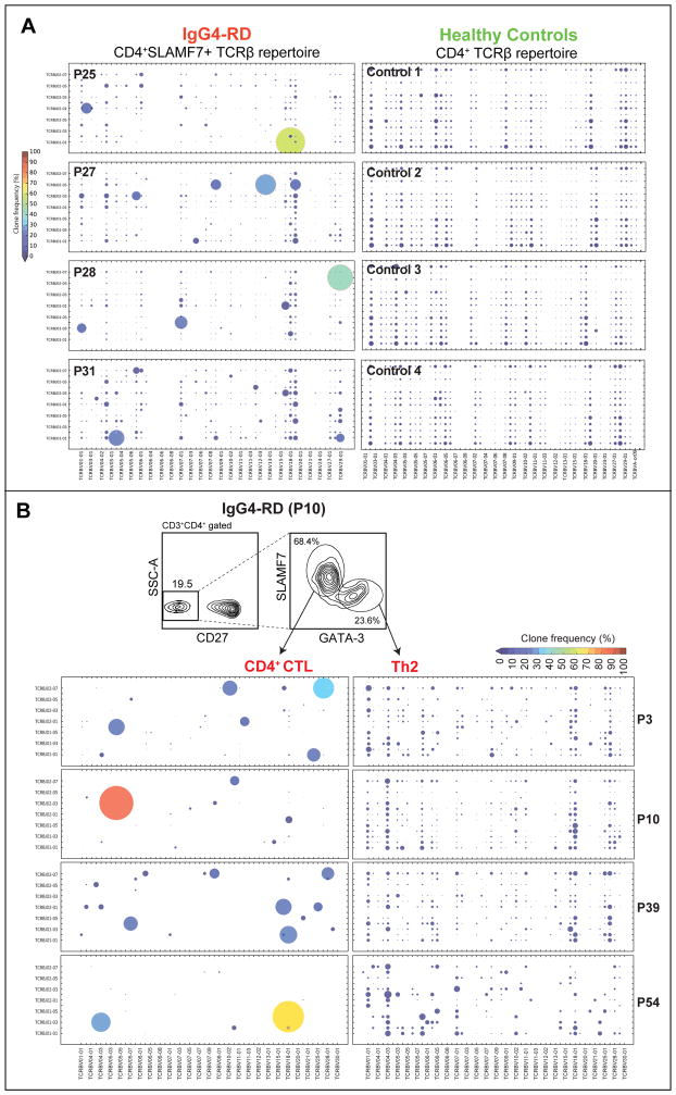
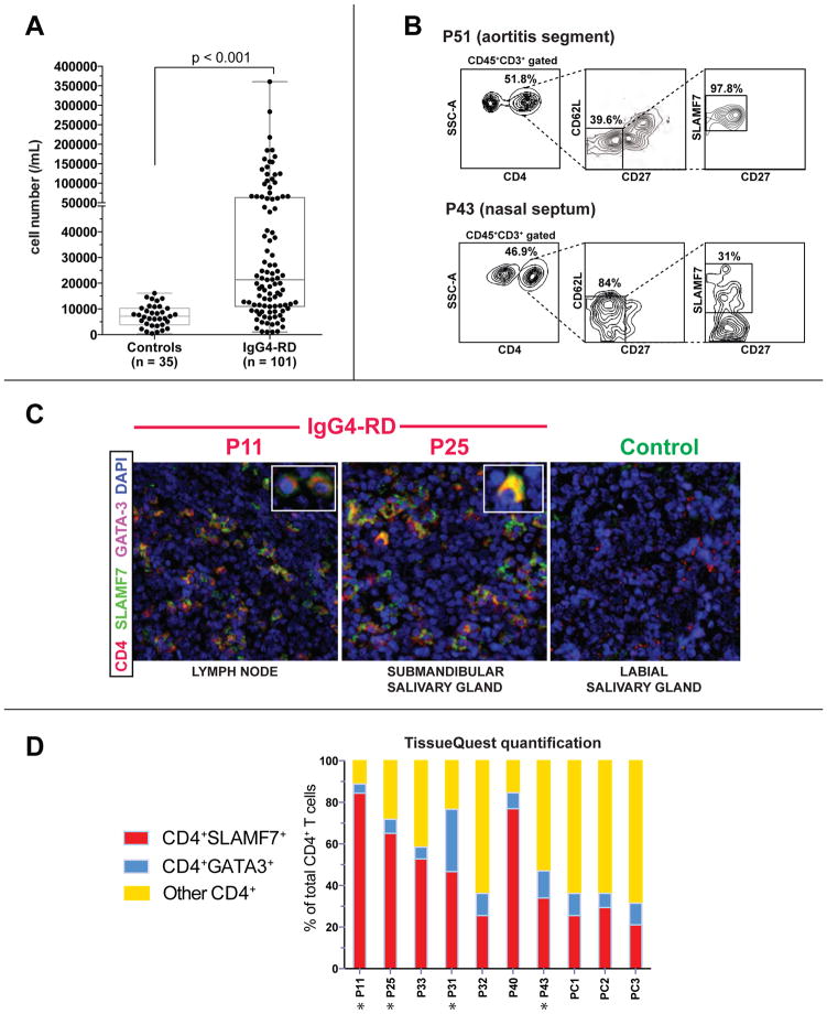
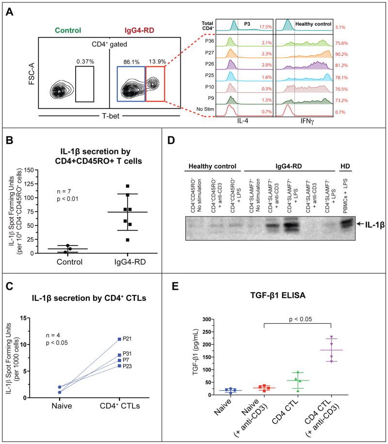
Methods: We used flow cytometry to identify CD4(+) effector/memory T cells in a cohort of 101 patients with IgG4-RD. These expanded cells were characterized by means of gene expression analysis and flow cytometry. Next-generation sequencing of the T-cell receptor β chain gene was performed on CD4(+)SLAMF7(+) cytotoxic T lymphocytes (CTLs) and CD4(+)GATA3(+) TH2 cells in a subset of patients to identify their clonality. Tissue infiltration by specific T cells was examined by using quantitative multicolor imaging.

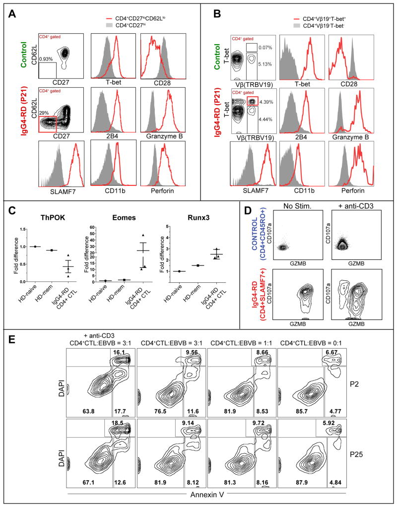
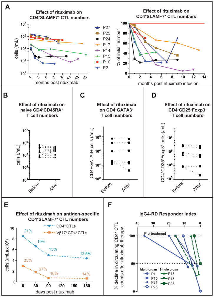
Results: CD4(+) effector/memory T cells with a cytolytic phenotype were expanded in patients with IgG4-RD. Next-generation sequencing revealed prominent clonal expansions of these CD4(+) CTLs but not CD4(+)GATA3(+) memory TH2 cells in patients with IgG4-RD. The dominant T cells infiltrating a range of inflamed IgG4-RD tissue sites were clonally expanded CD4(+) CTLs that expressed SLAMF7, granzyme A, IL-1β, and TGF-β1. Clinical remission induced by rituximab-mediated B-cell depletion was associated with a reduction in numbers of disease-associated CD4(+) CTLs.

Conclusions: IgG4-RD is prominently linked to clonally expanded IL-1β- and TGF-β1-secreting CD4(+) CTLs in both peripheral blood and inflammatory tissue lesions. These active, terminally differentiated, cytokine-secreting effector CD4(+) T cells are now linked to a human disease characterized by chronic inflammation and fibrosis.

Keywords: CD4(+) cytotoxic T cells; Fibrosis; IL-1β; IgG(4); IgG(4)-related disease; T(H)2 cells; rituximab.


Conflict of interest statement

The authors declare that they have no relevant conflict of interest.

Figures

Figure 1
Figure 1. Expansions of TEM cells with a cytolytic phenotype in IgG4-RD
(A) CD4+CD62LloCD27lo TEM cells in two non-atopic (P2 & P19) two atopic (P12 & P25) IgG4-RD patients and 2 representative healthy controls. (B) CD4+CD45RO+CD62LloCD27lo TEM cells in peripheral blood of IgG4-RD subjects. Boxplot displays the 25th to 75th percentiles. P values are based on the Mann-Whitney test. (C) Heat map depicting differentially expressed immune-related genes in TEM cells from four IgG4-RD patients compared to CD4+CD45RO+ T cells from four healthy controls. Upregulated genes representing modified Th1, cytolytic and myeloid signatures are highlighted.
Figure 2
Figure 2. Expanded TEM cells in IgG4-RD subjects exhibit a functional cytolytic profile
(A & B) Key hits from the gene expression analysis from Fig. 1 were validated using flow cytometry in TEM cells (A) and using TCR Vβ-specific antibodies (B). (C) Levels of ThPOK and Runx3 in CD4+SLAMF7+ CTLs from IgG4-RD subjects and CD4+CD45RO+ T cells in healthy controls. Error bars show SEM. P values are based on the Mann-Whitney test. (D) Granzyme B and CD107a staining on CD4+ CTLs from an IgG4-RD patient, before and after stimulation with anti-CD3 . (E) Cytotoxicity of in vitro expanded CD4+ CTLs derived from two subjects (P2 and P25) against an allogeneic EBV-transformed B cell target cell line, measured after 12 hours of co-culture with or without anti-CD3 (10 μg/mL) at varying CD4+ CTL: target ratios. The data are representative of two separate experiments done on 4 patients.
Figure 3
Figure 3. The TCR Vβ repertoire of expanded TEM cells in IgG4-RD
(A) TCRβ repertoire of the expanded circulating TEM subset in 4 IgG4-RD subjects (P31, P28, P27 and P25) and from total CD4+ T cells from four healthy controls, represented as bubble charts, where the size and color corresponds to the frequency of the observed Vβ-Jβ rearrangements. (B) TCRβ repertoire of expanded circulating CD4+GATA-3+ TH2 cells in four IgG4-RD subjects with an atopic history (P3, P10, P39 & P54), contrasted with the expanded CD4+SLAMF7+ CTLs from the same individuals. See legend to Fig.3A for details.
Figure 4
Figure 4. Expansion of CD4+SLAMF7+ CTLs in tissue lesions of IgG4-RD subjects
(A) Expansion of CD4+SLAMF7+ CTLs in 101 IgG4-RD subjects. (B) CD4+CD62LloCD27loSLAMF7+ CTLs in the aortic wall of a subject with IgG4-related aortitis (P51) and the involved nasal septum of a subject with IgG4-RD (P43). (C) Immunofluorescence staining of CD4+SLAMF7+ CTLs and CD4+GATA-3+ Th2 cells in the affected tissues of IgG4-RD subjects (lymph node biopsy from P11 and salivary gland biopsy from P25). CD4 (red), DAPI (blue), GATA-3 (magenta) and SLAMF7 (green) staining are shown. (D) Quantification of CD4+SLAMF7+, CD4+GATA-3+ and CD4+SLAMF7-GATA-3- cells in tissue biopsies from 10 IgG4-RD patients (atopic patients are marked with asterisks).
Figure 5
Figure 5. Disease associated CD4+ CTLs secrete IFNγ, IL-1β and TGF-β1 upon in vitro restimulation
(A) Intracellular staining for IFN-γ and IL-4 in restimulated CD4+ CTLs from seven IgG4-RD patients. The empty histogram (red) depicts an unstimulated control. (B & C) ELISPOT detection of the frequency of IL-1β producers among re-stimulated CD4+CD45RO+ T cells from seven IgG4-RD subjects compared to healthy donors (error bars show SEM, unpaired t-test) (A) and CD4+ CTLs from four IgG4-RD subjects compared to naïve CD4+CD45RA+ T cells from the same individuals (p< 0.05, paired t-test) (B). (D) Western Blot detection of IL-1β from culture supernatants of in vitro expanded T cells, maintained in IL-2 for two weeks (10 ng/mL). Supernatants from CD4+CD45RO+ T cells from a healthy donor and CD4+CD45RO+SLAMF7+ or CD4+CD45RO+SLAMF7 T cells from an IgG4-RD subject were used without any stimulation (US), with 5 μg/mL LPS or 3μg/mL anti-CD3. LPS stimulated PBMCs from a healthy donor were used as a positive control. (E) Detection of TGF-β1 production among naïve CD4+ cells and CD4+SLAMF7+ CTLs from four IgG4-RD subjects by ELISA, with or without in vitro re-stimulation (with 3 μg/mL anti-CD3). Error bars show SEM. P values are based on the Mann-Whitney test.
Figure 6
Figure 6. Expansion of IL-1β and TGF-β1 secreting CD4+ CTLs in IgG4-RD lesions
(A) Immunofluorescence staining of IL-1β-producing CD4+ SLAMF7+ cells in the tissues of 2 IgG4-RD subjects (lymph node biopsy from P11 and salivary gland biopsy from P25) and a healthy control subject (labial salivary gland biopsy). CD4 (red), SLAMF7 (green) DAPI (blue) and IL-1β (white). (B) Immunofluorescence staining of TGF-β1-producing CD4+ SLAMF7+ cells in the tissues of 2 IgG4-DS and 2 Sjögren’s syndrome subjects (submandibular salivary gland biopsies). CD4 (red), Granzyme A (green) DAPI (blue) and IL-1β (cyan) staining are shown.
Figure 7
Figure 7. Rituximab-mediated depletion of CD4+ CTLs
(A) Decline in CD4+ SLAMF7+ CTLs (n = 8 subjects) over 6–14 months following rituximab infusion shown as a change in cell counts and proportion. (B) Effect of rituximab on CD4+ CD45RA+ naïve T cell counts in the peripheral blood of IgG4-RD patients. (C & D) Effect of rituximab on CD4+GATA-3+ TH2 cells and CD4+CD25+Foxp3+ cells in the peripheral blood of IgG4-RD patients 90–120 days after rituximab therapy. (E) Decline in the number and proportion of an expanded CD4+ CTL clone tracked using a TCR-Vβ specific antibody following rituximab therapy in an IgG4-RD subject (P25). (F) Decline in circulating CD4+ CTL numbers at day 70–95 following rituximab therapy (normalized to the pretreatment levels) is plotted against the IgG4-RD Responder Index, a clinical measure of disease activity.

References

    1. Stone JH, Zen Y, Deshpande V. IgG4-related disease. N Engl J Med. 2012;366:539–51. - PubMed
    1. Mahajan VS, Mattoo H, Deshpande V, Pillai SS, Stone JH. IgG4-Related Disease. Annu Rev Pathol. 2014;9:315–47. - PubMed
    1. Kamisawa T, Zen Y, Pillai S, Stone JH. IgG4-related disease. Lancet. 2015;385:1460–71. - PubMed
    1. Bindon CI, Hale G, Bruggemann M, Waldmann H. Human monoclonal IgG isotypes differ in complement activating function at the level of C4 as well as C1q. J Exp Med. 1988;168:127–42. - PMC - PubMed
    1. Bruhns P, Iannascoli B, England P, Mancardi DA, Fernandez N, Jorieux S, et al. Specificity and affinity of human Fcgamma receptors and their polymorphic variants for human IgG subclasses. Blood. 2009;113:3716–25. - PubMed

Publication types