Review
. 2016 Aug;105(8):463-75.
doi: 10.1002/bip.22832.

Review: Translational GTPases

Affiliations
Review

Review: Translational GTPases

Cristina Maracci et al. Biopolymers. 2016 Aug.

Abstract

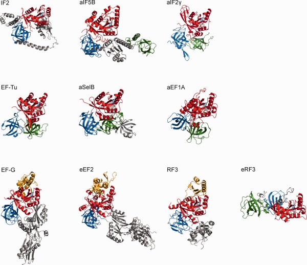
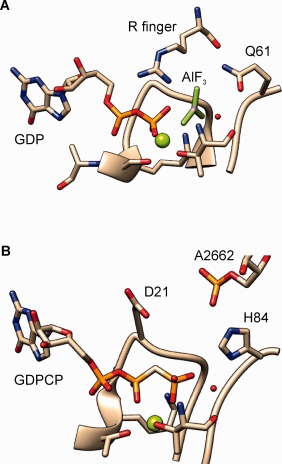
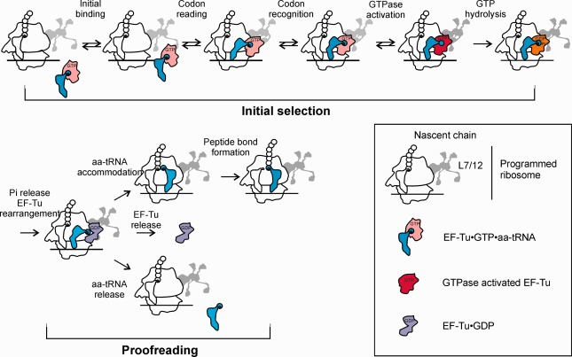
Translational GTPases (trGTPases) play key roles in facilitating protein synthesis on the ribosome. Despite the high degree of evolutionary conservation in the sequences of their GTP-binding domains, the rates of GTP hydrolysis and nucleotide exchange vary broadly between different trGTPases. EF-Tu, one of the best-characterized model G proteins, evolved an exceptionally rapid and tightly regulated GTPase activity, which ensures rapid and accurate incorporation of amino acids into the nascent chain. Other trGTPases instead use the energy of GTP hydrolysis to promote movement or to ensure the forward commitment of translation reactions. Recent data suggest the GTPase mechanism of EF-Tu and provide an insight in the catalysis of GTP hydrolysis by its unusual activator, the ribosome. Here we summarize these advances in understanding the functional cycle and the regulation of trGTPases, stimulated by the elucidation of their structures on the ribosome and the progress in dissecting the reaction mechanism of GTPases. © 2016 Wiley Periodicals, Inc. Biopolymers 105: 463-475, 2016.

Keywords: EF-Tu; GTP hydrolysis; decoding; ribosome; tRNA; translation.


Figures

Figure 1
Figure 1
Structural overview of trGTPases. Crystal structures of trGTPases (PDB IDs: IF2 (4KJZ); aIF5B (1G7S); aIF2γ (1KK3); EF‐Tu (1EFC); aSelB (4AC9); aEF1A (1SKQ); EF‐G (2EFG); eEF2 (1N0U); RF3 (2HSE); eRF3 (1R5N)) in the complex with GDP (except for eEF2, which is shown in the apo‐form) were aligned on the P‐loop and GDP to highlight the structural conservation of the nucleotide binding (red) and the adjacent β‐barrel domains (blue and green, respectively). The G’ extension is represented in orange, the GDP molecule in cyan. Additional domains specific for each trGTPase are represented in gray.
Figure 2
Figure 2
trGTPases bind to the GAC center of the ribosome. Structure of the complex EF‐Tu·GDPCP·aa‐tRNA trapped on the programmed ribosome before GTP hydrolysis.20 In (a), the ribosome elements constituting the GAC are highlighted. The color code for EF‐Tu is the same as in Figure 1; the A/T aa‐tRNA is shown in magenta, the P‐site tRNA in purple, the E‐site tRNA in dark purple. (b) Close‐up view of the nucleotide binding pocket of EF‐Tu: the residues important for catalysis of GTP hydrolysis are shown as sticks; the catalytic water and the Mg2+ ion are shown in red and green, respectively (PDB: 2XQD and 2XQE).
Figure 3
Figure 3
Nucleotide exchange mechanism in EF‐Tu. Schematic of the nucleotide exchange pathway catalyzed by EF‐Ts. Binding of EF‐Ts to EF‐Tu–GDP causes dissociation of the Mg2+ ion, which initiates GDP dissociation. GTP is then loaded due to its higher cellular concentration compared to GDP. The change in the conformation into the GTP‐bound form occurs upon rebinding of Mg2+ and causes the dissociation of EF‐Ts.23, 40
Figure 4
Figure 4
Models for GTP hydrolysis of EF‐Tu on the ribosome. (a) General base mechanism; (b) substrate assisted pathway; (c) Two‐dimensional More O'Ferral‐Jencks plot for an ANDN reaction. The reaction coordinate proceeds from the lower left (ground state, GS) to the upper right corner (product state, PS). Bond cleavage and bond formation occur along the x and y axes, respectively. The TS can be located anywhere in the area, indicating that GTP hydrolysis can proceed via a continuum of reaction pathways. The farthermost cases of AN + DN and DN + AN will have additional intermediates in the upper left and lower right corners, respectively. Adapted from Ref. 65.
Figure 5
Figure 5
The active site of Ras and EF‐Tu in complex with the GAP. (a) Structure of the transition state of GTP hydrolysis in H‐Ras in complex with the RasGAP (PDB: 1WQ1) showing the Arg‐finger reaching the β‐γ bridging oxygen of the nucleotide; (b) Structure of the active site of EF‐Tu bound to the programmed ribosome in the pre‐hydrolysis state (PDB: 2XQD and 2XQE). The nucleophilic water molecule is highlighted in red, the Mg2+ in green.
Figure 6
Figure 6
EF‐Tu function during decoding on the ribosome. EF‐Tu‐dependent selection of cognate aa‐tRNA and rejection of aa‐tRNAs that do not match the A‐site codon is achieved in two selection steps separated by GTP hydrolysis by EF‐Tu. Shown and individual kinetically‐resolved steps identified by rapid kinetics and smFRET approaches. Adapted from Ref. 112.

References

    1. Bourne, H. R. ; Sanders, D. A. ; McCormick, F. Nature 1991, 349, 117–127. - PubMed
    1. Wittinghofer, A. ; Vetter, I. R. Annual Rev Biochem 2011, 80, 943–971. - PubMed
    1. Hilgenfeld, R. Curr Opin Struct Biol 1995, 5, 810–817. - PubMed
    1. Cherfils, J. ; Zeghouf, M. Physiol Rev 2013, 93, 269–309. - PubMed
    1. Bos, J. L. ; Rehmann, H. ; Wittinghofer, A. Cell 2007, 129, 865–877. - PubMed

Substances

LinkOut - more resources