Monocyte Mitochondrial Function in Calcium Oxalate Stone Formers
- PMID: 26972146
- PMCID: PMC4914421
- DOI: 10.1016/j.urology.2016.03.004
Monocyte Mitochondrial Function in Calcium Oxalate Stone Formers
Abstract
Objective: To investigate whether mitochondrial function is altered in circulating immune cells from calcium oxalate (CaOx) stone formers compared to healthy subjects.
Materials and methods: Adult healthy subjects (n = 18) and CaOx stone formers (n = 12) were included in a pilot study. Data collection included demographic and clinical values from electronic medical records. Bioenergetic function was assessed in monocytes, lymphocytes, and platelets isolated from blood samples using the Seahorse XF96 Analyzer. Plasma interleukin-6 (IL-6) was measured using enzyme-linked immunosorbent assay.
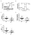
Results: All participants were age matched (44.5 ± 3.0 years for healthy subjects vs 42.3 ± 4.8 years for CaOx stone formers, P = .6905). CaOx stone formers did not have urinary tract infection, ureteral stones, or obstructing renal stones. Monocyte mitochondrial function was decreased in CaOx stone formers compared to healthy subjects. Specifically, mitochondrial maximal respiration (P = .0011) and reserve capacity (P < .0001) were significantly lower. In contrast, lymphocyte and platelet mitochondrial function was similar between the 2 groups. The bioenergetic health index, an integrated value of mitochondrial function, was significantly lower in monocytes from CaOx stone formers compared to healthy subjects (P = .0041). Lastly, plasma IL-6 levels were significantly increased (P = .0324).
Conclusion: The present pilot study shows that CaOx stone formers have decreased monocyte mitochondrial function. Plasma IL-6 was also increased in this cohort. These data suggest that impaired monocyte mitochondrial function and inflammation may be linked to CaOx kidney stone formation. Further studies are needed to confirm these findings in a larger cohort of patients.
Copyright © 2016 Elsevier Inc. All rights reserved.
Conflict of interest statement
Figures


Comment in
-
Re: Monocyte Mitochondrial Function in Calcium Oxalate Stone Formers.J Urol. 2016 Oct;196(4):1170-1. doi: 10.1016/j.juro.2016.07.006. Epub 2016 Jul 13. J Urol. 2016. PMID: 27628803 No abstract available.
Similar articles
-
Clinical and biochemical profile of patients with "pure" uric acid nephrolithiasis compared with "pure" calcium oxalate stone formers.Urol Res. 2007 Oct;35(5):247-51. doi: 10.1007/s00240-007-0109-1. Epub 2007 Sep 6. Urol Res. 2007. PMID: 17786420
-
Clinical and Metabolic Correlates of Calcium Oxalate Stone Subtypes: Implications for Etiology and Management.J Endourol. 2019 Sep;33(9):755-760. doi: 10.1089/end.2019.0245. Epub 2019 Jul 31. J Endourol. 2019. PMID: 31154910
-
Metabolic syndrome and the risk of calcium stones.Nephrol Dial Transplant. 2012 Aug;27(8):3201-9. doi: 10.1093/ndt/gfr703. Epub 2012 Jan 13. Nephrol Dial Transplant. 2012. PMID: 22247230 Free PMC article.
-
Dietary treatment of urinary risk factors for renal stone formation. A review of CLU Working Group.Arch Ital Urol Androl. 2015 Jul 7;87(2):105-20. doi: 10.4081/aiua.2015.2.105. Arch Ital Urol Androl. 2015. PMID: 26150027 Review.
-
Nutritional aspects of stone disease.Endocrinol Metab Clin North Am. 2002 Dec;31(4):1017-30, ix-x. doi: 10.1016/s0889-8529(02)00029-4. Endocrinol Metab Clin North Am. 2002. PMID: 12474643 Review.
Cited by
-
Mitochondrial Dysfunction and Kidney Stone Disease.Front Physiol. 2020 Oct 20;11:566506. doi: 10.3389/fphys.2020.566506. eCollection 2020. Front Physiol. 2020. PMID: 33192563 Free PMC article. Review.
-
Oxalate Alters Cellular Bioenergetics, Redox Homeostasis, Antibacterial Response, and Immune Response in Macrophages.Front Immunol. 2021 Oct 21;12:694865. doi: 10.3389/fimmu.2021.694865. eCollection 2021. Front Immunol. 2021. PMID: 34745086 Free PMC article.
-
Hydroxyproline stimulates inflammation and reprograms macrophage signaling in a rat kidney stone model.Biochim Biophys Acta Mol Basis Dis. 2022 Sep 1;1868(9):166442. doi: 10.1016/j.bbadis.2022.166442. Epub 2022 May 10. Biochim Biophys Acta Mol Basis Dis. 2022. PMID: 35562038 Free PMC article.
-
Overview of methods that determine mitochondrial function in human disease.Metabolism. 2025 Sep;170:156300. doi: 10.1016/j.metabol.2025.156300. Epub 2025 May 17. Metabolism. 2025. PMID: 40389059 Free PMC article. Review.
-
Oxalate (dys)Metabolism: Person-to-Person Variability, Kidney and Cardiometabolic Toxicity.Genes (Basel). 2023 Aug 29;14(9):1719. doi: 10.3390/genes14091719. Genes (Basel). 2023. PMID: 37761859 Free PMC article. Review.
References
-
- Shoag J, Halpern J, Goldfarb DS, Eisner BH. Risk of chronic and end stage kidney disease in patients with nephrolithiasis. J Urol. 2014;192:1440–5. - PubMed
-
- Holmes RP, Assimos DG. The impact of dietary oxalate on kidney stone formation. Urol Res. 2004;32:311–6. - PubMed
-
- Goodman HO, Brommage R, Assimos DG, Holmes RP. Genes in idiopathic calcium oxalate stone disease. World J Urol. 1997;15:186–94. - PubMed
MeSH terms
Substances
Grants and funding
LinkOut - more resources
Full Text Sources
Other Literature Sources

