

Immunohistochemical expression of heparanase isoforms and syndecan-1 proteins in colorectal adenomas
- PMID: 26972718
- PMCID: PMC4800254
- DOI: 10.4081/ejh.2016.2590
Immunohistochemical expression of heparanase isoforms and syndecan-1 proteins in colorectal adenomas
Abstract
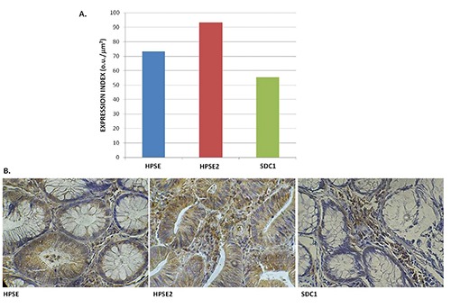
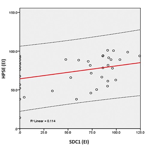
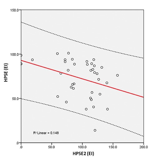
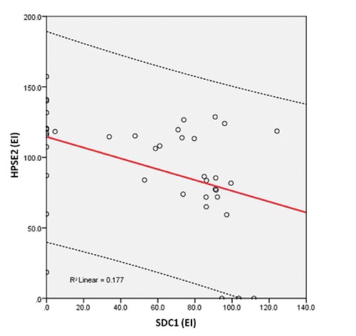
The proteoglycan syndecan-1 and the endoglucuronidases heparanase-1 and heparanase-2 are involved in molecular pathways that deregulate cell adhesion during carcinogenesis. Few studies have examined the expression of syndecan-1, heparanase-1 and mainly heparanase-2 proteins in non-neoplastic and neoplastic human colorectal adenoma tissues. The aim of this study was to analyze the correlation among the heparanase isoforms and the syndecan-1 proteins through immunohistochemical expression in the tissue of colorectal adenomas. Primary anti-human polyclonal anti-HPSE and anti-HPSE2 antibodies and primary anti-human monoclonal anti-SDC1 antibody were used in the immunohistochemical study. The expressions of heparanase-1 and heparanase-2 proteins were determined in tissue samples from 65 colorectal adenomas; the expression of syndecan-1 protein was obtained from 39 (60%) patients. The histological type of adenoma was tubular in 44 (67.7%) patients and tubular-villous in 21 (32.3%); there were no villous adenomas. The polyps were <1.0 cm in size in 54 (83.1%) patients and ≥1.0 cm in 11 (16.9%). The images were quantified by digital counter with a computer program for this purpose. The expression index represented the relationship between the intensity expression and the percentage of positively stained cells. The results showed that the average of heparanase-1, heparanase-2 and syndecan-1 expression index was 73.29 o.u./µm², 93.34 o.u./µm², and 55.29 o.u./µm², respectively. The correlation between the heparanase-1 and syndecan-1 expression index was positive (R=0.034) and significant (P=0.035). There was a negative (R= -0.384) and significant (P=0.016) correlation between the expression index of heparanase-1 and heparanase-2. A negative (R= -0.421) and significant (P=0.008) correlation between the expression index of heparanase-2 and syndecan-1 was found. We concluded that in colorectal adenomas, the heparanase-1 does not participate in syndecan-1 degradation; the heparanase-2 does not stimulate syndecan-1 degradation by the action of heparanase-1, and the heparanase-2 may be involved in the modulation of the heparanase-1 activity.
Conflict of interest statement
Conflict of interest: the authors declare no conflict of interest.
Figures
References
Publication types
MeSH terms
Substances
LinkOut - more resources
Full Text Sources
Other Literature Sources
Medical
Miscellaneous
