. 2016 Mar 14:6:23010.
doi: 10.1038/srep23010.

Deletion of interleukin-6 alleviated interstitial fibrosis in streptozotocin-induced diabetic cardiomyopathy of mice through affecting TGFβ1 and miR-29 pathways

Affiliations

Deletion of interleukin-6 alleviated interstitial fibrosis in streptozotocin-induced diabetic cardiomyopathy of mice through affecting TGFβ1 and miR-29 pathways

Yang Zhang et al. Sci Rep. .

Abstract

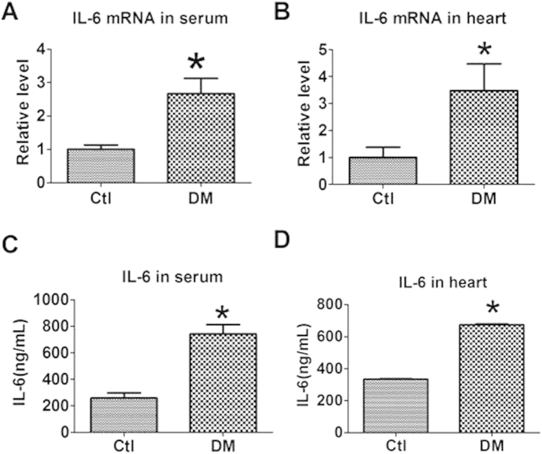
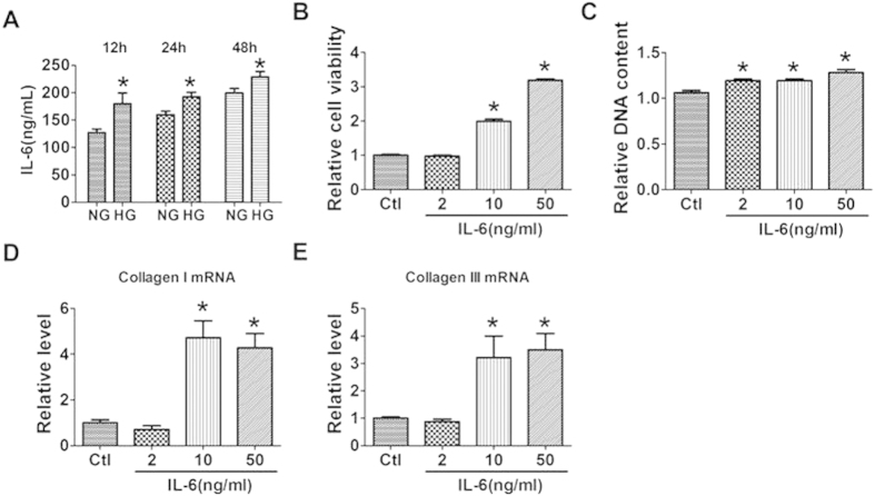
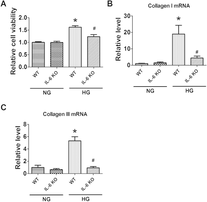
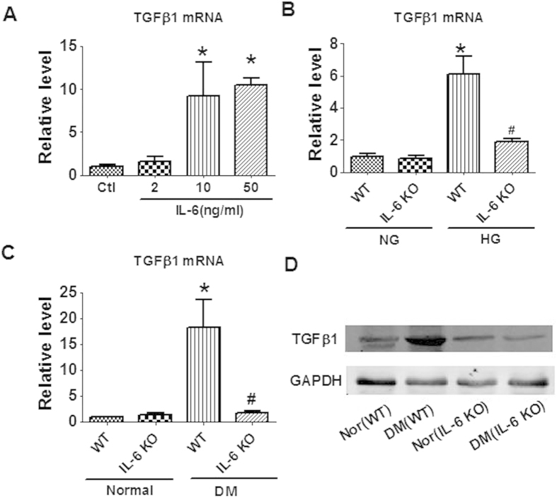
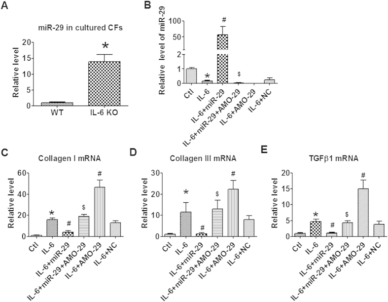
Interleukin 6 (IL-6) has been shown to be an important regulator of cardiac interstitial fibrosis. In this study, we explored the role of interleukin-6 in the development of diabetic cardiomyopathy and the underlying mechanisms. Cardiac function of IL-6 knockout mice was significantly improved and interstitial fibrosis was apparently alleviated in comparison with wildtype (WT) diabetic mice induced by streptozotocin (STZ). Treatment with IL-6 significantly promoted the proliferation and collagen production of cultured cardiac fibroblasts (CFs). High glucose treatment increased collagen production, which were mitigated in CFs from IL-6 KO mice. Moreover, IL-6 knockout alleviated the up-regulation of TGFβ1 in diabetic hearts of mice and cultured CFs treated with high glucose or IL-6. Furthermore, the expression of miR-29 reduced upon IL-6 treatment, while increased in IL-6 KO hearts. Overexpression of miR-29 blocked the pro-fibrotic effects of IL-6 on cultured CFs. In summary, deletion of IL-6 is able to mitigate myocardial fibrosis and improve cardiac function of diabetic mice. The mechanism involves the regulation of IL-6 on TGFβ1 and miR-29 pathway. This study indicates the therapeutic potential of IL-6 suppression on diabetic cardiomyopathy disease associated with fibrosis.


Figures

Figure 1
Figure 1. Level of IL-6 in the serum and heart of mice with diabetes mellitus (DM).
(A,B) IL-6 mRNA level in the serum and heart by qRT-PCR. (C,D) IL-6 level in the serum and heart by Immunoassay. Data are expressed as mean ± SEM. N = 3. Ctl, control. *P < 0.05 vs Ctl.
Figure 2
Figure 2. Effect of IL-6 knockout on cardiac function of diabetic mice by echocardiography.
(A) Eject fraction (EF). (B) Fractional shortening (FS). (C) E/A ratio. (D) Left ventricular mass (LV Mass). WT, wild-type; IL-6 KO, IL-6 knockout; DM, Diabetes mellitus. Data are expressed as mean ± SEM. N = 7 or 8. *P < 0.05 vs normal WT, #P < 0.05 vs DM WT.
Figure 3
Figure 3. Effect of IL-6 knockout on interstitial fibrosis of diabetic mice.
(A,B) mRNA levels of collagen I and collagen III. WT, wild-type; IL-6 KO, IL-6 knockout; DM, Diabetes mellitus. Data are expressed as mean ± SEM. n = 5. *P < 0.05 vs normal WT, #P < 0.05 vs DM WT. (C) Protein expression of collagen I and collagen III by western blot. WT, wild-type; IL-6 KO, IL-6 knockout; Nor, Normal; DM, Diabetes mellitus. (D) Deposition of collagen in cardiac tissue by Masson’s staining. Scale bar = 100 μm.
Figure 4
Figure 4. Effects of interleukin-6 (IL-6) on the proliferation of cultured cardiac fibroblasts (CFs).
(A) Effects of glucose treatment on the production of IL-6 in CFs by immunoassay. (B,C) Effects of IL-6 treatment on the viability and proliferation of CFs by MTT assay and DNA incorporation assay. (D,E) mRNA levels of collagen I and collagen III in CFs treated with IL-6. Ctl, Control. NG, normal glucose; HG, high glucose. Ctl, control. Data are expressed as mean ± SEM. N = 4. *P < 0.05 vs NG or Ctl.
Figure 5
Figure 5. Effects of IL-6 knockout (IL-6 KO) on the proliferation of cultured cardiac fibroblasts (CFs) stimulated with high glucose (HG).
(A) Cell viability evaluated by MTT assay. (B,C) mRNA levels of collagen I and collagen III. NG, Normal glucose. WT, Wild-type. Data are expressed as mean ± SEM. N = 4. *P < 0.05 vs NG WT, #P < 0.05 vs HG WT.
Figure 6
Figure 6. Effects of IL-6 on TGFβ1 production.
(A) Application of IL-6 increased the production of TGFβ1 in cultured cardiac fibroblasts (CFs) from wild type (WT) mice. (B) Knockout of IL-6 inhibited the production of TGFβ1 induced by high glucose (HG) in cultured CFs. (C) Knockout of IL-6 inhibited the production of TGFβ1 in the heart of diabetic mice. (D) Protein expression of TGFβ1 by western blot. Ctl, control; NG, Normal glucose; Nor, Normal; DM, Diabetes mellitus; WT, Wild-type. Data are expressed as mean ± SEM. N = 4. *P < 0.05 vs WT or Ctl; #P < 0.05 vs WT (HG or DM).
Figure 7
Figure 7. miR-29 on IL-6 induced collagenI and TGFB1 production in cultured CFs.
(A) miR-29 levels in cultured cardiac fibroblasts (CFs) isolated from wild type (WT) and IL-6 knockout (IL-6 KO) mice. (B) miR-29 levels after IL-6 stimulation and exogenous miR-29/AMO-29 treatment. (CE) mRNA levels of collagen I, collagen III and TGFβ1 in cultured CFs treated with IL-6 and miR-29/AMO-29. Ctl, Control; NC, Negative control. Data are expressed as mean ± SEM. N = 4. *P < 0.05 vs WT or Ctl; #P < 0.05 vs IL-6; $P < 0.05 vs IL-6 + miR-29.
Figure 8
Figure 8
Effect of high glucose and IL-6 knockout on the expression of miR-29 in cultured CFs (A) and cardiac tissue (B) Data are expressed as mean ± SEM. N = 4. *P < 0.05 vs NG or WT; #P < 0.05 vs HG or DM.
Figure 9
Figure 9. A schematic graph on the signaling pathway of the regulation of IL-6 on cardiac fibrosis and DCM.DCM, diabetic cardiomyopathy.
Blue line with hammer head for “inhibition”; red line with arrow head for “stimulation”.

References

    1. Pappachan J. M., Varughese G. I., Sriraman R. & Arunagirinathan G. Diabetic cardiomyopathy: Pathophysiology, diagnostic evaluation and management. World J Diabetes 4, 177–189 (2013). - PMC - PubMed
    1. Galderisi M. Diastolic dysfunction and diabetic cardiomyopathy: evaluation by Doppler echocardiography. J Am Coll Cardiol 48, 1548–1551 (2006). - PubMed
    1. Goyal B. R. & Mehta A. A. Diabetic cardiomyopathy: pathophysiological mechanisms and cardiac dysfuntion. Hum Exp Toxicol 32, 571–590 (2013). - PubMed
    1. Li Z., Abdullah C. S. & Jin Z. Q. Inhibition of PKC-theta preserves cardiac function and reduces fibrosis in streptozotocin-induced diabetic cardiomyopathy. Br J Pharmacol 171, 2913–2924 (2014). - PMC - PubMed
    1. Wang W. K. et al. Inhibition of high-mobility group box 1 improves myocardial fibrosis and dysfunction in diabetic cardiomyopathy. Int J Cardiol 172, 202–212 (2014). - PubMed

Publication types

MeSH terms