. 2016 Jul 1:103:167-74.
doi: 10.1016/j.ymeth.2016.03.003. Epub 2016 Mar 10.

In vitro RNA SELEX for the generation of chemically-optimized therapeutic RNA drugs

Affiliations

In vitro RNA SELEX for the generation of chemically-optimized therapeutic RNA drugs

Kevin T Urak et al. Methods. .

Abstract

Aptamers are single-stranded DNA or RNA oligonucleotides that can bind with exquisitely high affinity and specificity to target molecules and are thus often referred to as 'nucleic acid' antibodies. Oligonucleotide aptamers are derived through a process of directed chemical evolution called SELEX (Systematic Evolution of Ligands by Exponential enrichment). This chemical equivalent of Darwinian evolution was first described in 1990 by Tuerk & Gold and Ellington & Szostak and has since yielded aptamers for a wide-range of applications, including biosensor technologies, in vitro diagnostics, biomarker discovery, and therapeutics. Since the inception of the original SELEX method, numerous modifications to the protocol have been described to fit the choice of target, specific conditions or applications. Technologies such as high-throughput sequencing methods and microfluidics have also been adapted for SELEX. In this chapter, we outline key steps in the SELEX process for enabling the rapid identification of RNA aptamers for in vivo applications. Specifically, we provide a detailed protocol for the selection of chemically-optimized RNA aptamers using the original in vitro SELEX methodology. In addition, methods for performing next-generation sequencing of the RNAs from each round of selection, based on Illumina sequencing technology, are discussed.

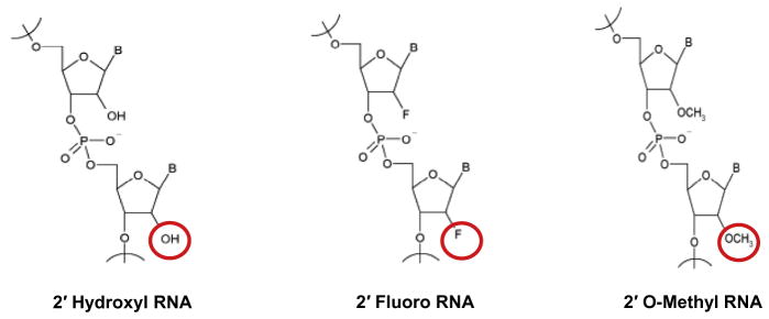
Keywords: 2′-Fluoro pyrimidines; Illumina sequencing; In vitro SELEX; Modified nucleotides; Mutant T7 RNA polymerase; Next-generation sequencing; RNA aptamers; Recombinant proteins.


Figures

Fig. 1
Fig. 1
Predicted tertiary structure of PSMA RNA aptamer A9g. The predicted tertiary structure (3D) of A9g was obtained as described in Rockey et al. (2011). The constructed PDB files were then processed using the Swiss PDBViewer to generate the 3D structure. This aptamer is chemically modified with 2′ fluoro chemistry for clinical applications. A9g is a non-competitive inhibitor of PSMA enzymatic activity. It inhibits PSMA-mediated migration and invasion of prostate cancer cells in culture and dissemination in a mouse model of metastatic bone disease.
Fig. 2
Fig. 2
Commonly used 2′-ribose modified nucleotides in aptamer selection experiments. The mutated T7 (Y639F) RNA polymerase is able to easily incorporate the 2′OH and 2′F modifications during selection, while the 2′Ome modification is normally incorporated synthetically after selection has occurred. (Modified nucleotides and aptamer libraries can be purchased from TriLink BioTechnologies, Inc. – http://www.trilinkbiotech.com/.)
Fig. 3
Fig. 3
Gel running apparatus (Fisher Biotech FB-VE20-1, left) and high-voltage power supply (Bio-Rad model 3000Xi, right).
Fig. 4
Fig. 4
Visual representation of UV shadowing. (A) Schematic of set-up for UV-shadowing of RNA. The denaturing urea-PAGE gel (shown in grey) is positions on top of the TLC plate (shown in white). UV light (purple) is shined on top of the gel. A shadow will appear in the place where the RNA aptamer is on the gel. The RNA aptamer runs between the top dye (xylene cyanol) and the dye front (bromophenol blue). (B) UV shadow of an RNA aptamer visualized using the TLC plate method. The RNA is run on a denaturing Urea-PAGE gel until the dye front reaches the bottom of the gel. The RNA band (shadow) can be easily excised using a clean razor blade and the RNA eluted from the gel in 1X TE buffer.

References

    1. Keefe AD, Pai S, Ellington A. Aptamers as therapeutics. Nat Rev Drug Discovery. 2010;9(7):537–550. - PMC - PubMed
    1. Diafa S, Hollenstein M. Generation of aptamers with an expanded chemical repertoire. Molecules. 2015;20(9):16643–16671. - PMC - PubMed
    1. Dassie JP, et al. Targeted inhibition of prostate cancer metastases with an RNA aptamer to prostate-specific membrane antigen. Mol Ther. 2014;22(11):1910–1922. - PMC - PubMed
    1. Rockey WM, et al. Rational truncation of an RNA aptamer to prostate-specific membrane antigen using computational structural modeling. Nucleic Acid Ther. 2011;21(5):299–314. - PMC - PubMed
    1. Ellington AD, Szostak JW. In vitro selection of RNA molecules that bind specific ligands. Nature. 1990;346(6287):818–822. - PubMed

Publication types

MeSH terms

Substances

LinkOut - more resources