. 2016 Jul;215(1 Suppl):S1-S46.
doi: 10.1016/j.ajog.2016.03.001. Epub 2016 Mar 10.

Placental origins of adverse pregnancy outcomes: potential molecular targets: an Executive Workshop Summary of the Eunice Kennedy Shriver National Institute of Child Health and Human Development

Affiliations

Placental origins of adverse pregnancy outcomes: potential molecular targets: an Executive Workshop Summary of the Eunice Kennedy Shriver National Institute of Child Health and Human Development

John V Ilekis et al. Am J Obstet Gynecol. 2016 Jul.

Abstract

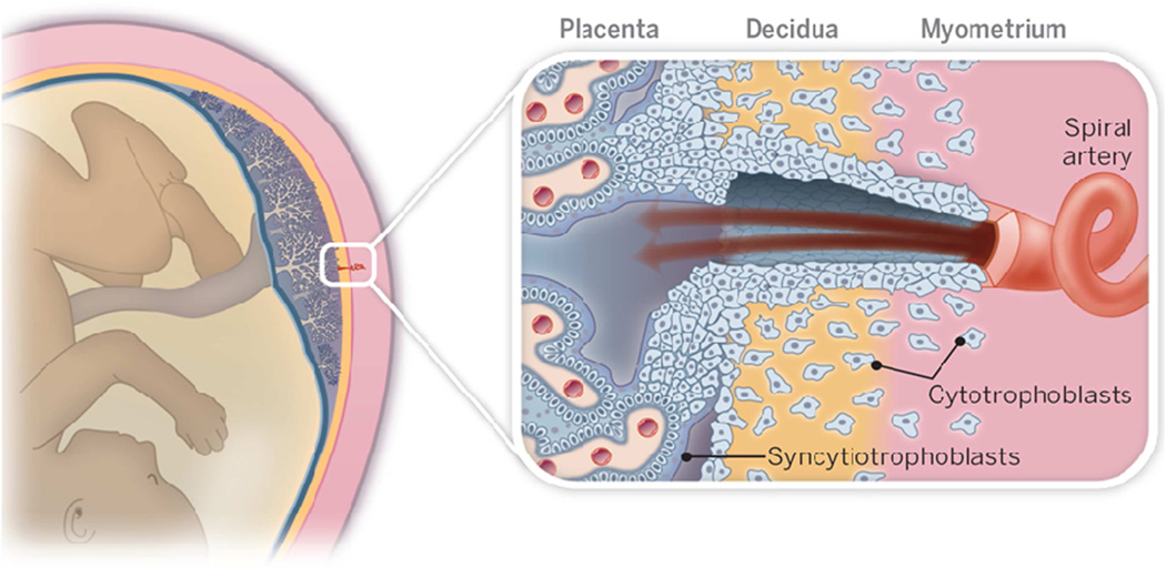
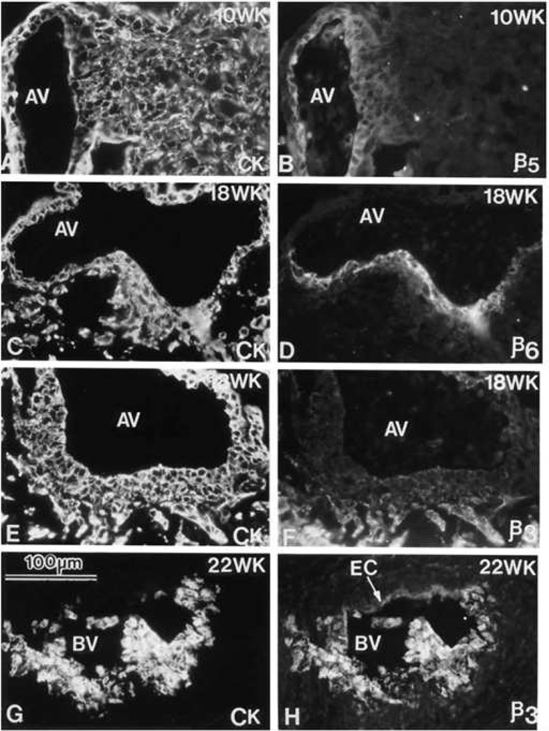
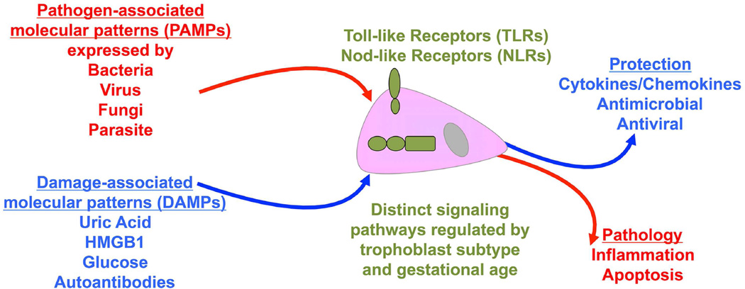
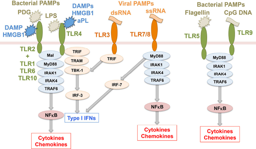
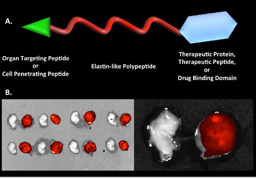
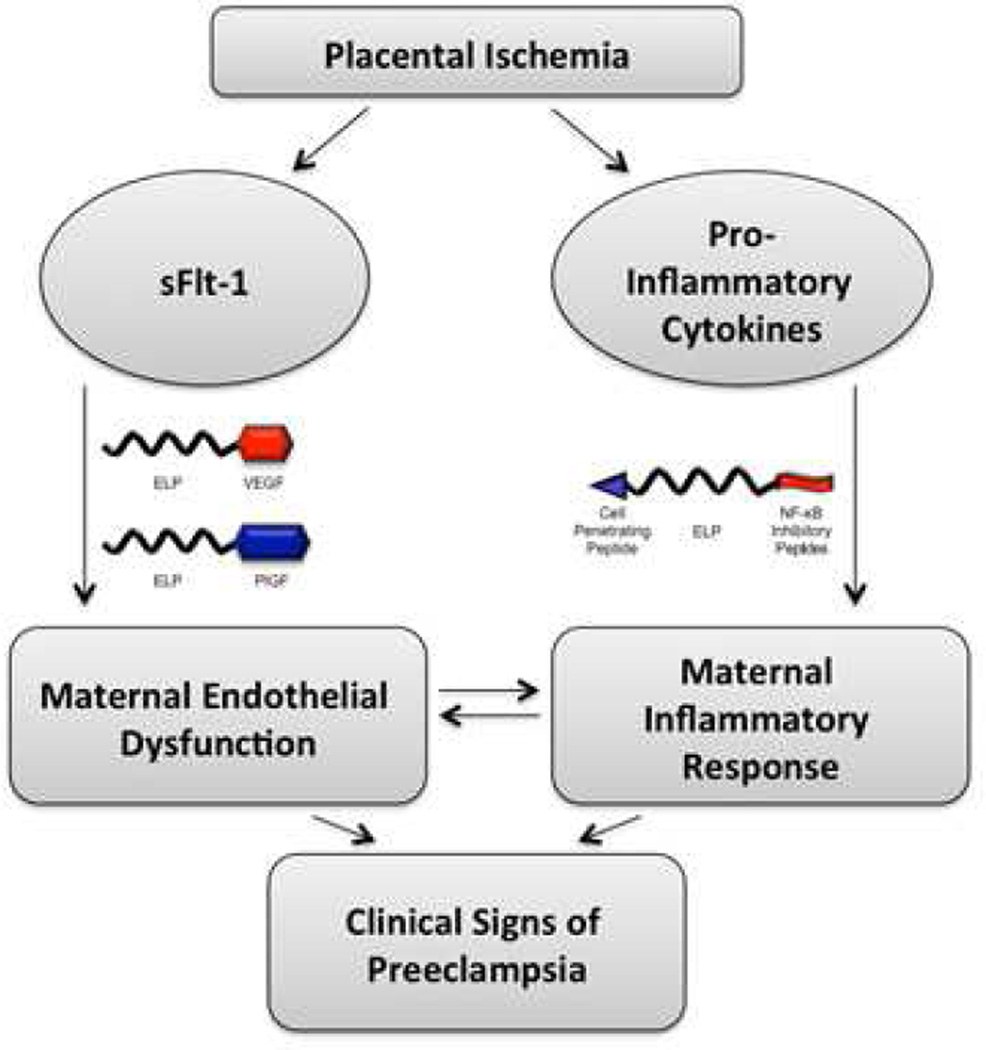
Although much progress is being made in understanding the molecular pathways in the placenta that are involved in the pathophysiology of pregnancy-related disorders, a significant gap exists in the utilization of this information for the development of new drug therapies to improve pregnancy outcome. On March 5-6, 2015, the Eunice Kennedy Shriver National Institute of Child Health and Human Development of the National Institutes of Health sponsored a 2-day workshop titled Placental Origins of Adverse Pregnancy Outcomes: Potential Molecular Targets to begin to address this gap. Particular emphasis was given to the identification of important molecular pathways that could serve as drug targets and the advantages and disadvantages of targeting these particular pathways. This article is a summary of the proceedings of that workshop. A broad number of topics were covered that ranged from basic placental biology to clinical trials. This included research in the basic biology of placentation, such as trophoblast migration and spiral artery remodeling, and trophoblast sensing and response to infectious and noninfectious agents. Research findings in these areas will be critical for the formulation of the development of future treatments and the development of therapies for the prevention of a number of pregnancy disorders of placental origin that include preeclampsia, fetal growth restriction, and uterine inflammation. Research was also presented that summarized ongoing clinical efforts in the United States and in Europe that has tested novel interventions for preeclampsia and fetal growth restriction, including agents such as oral arginine supplementation, sildenafil, pravastatin, gene therapy with virally delivered vascular endothelial growth factor, and oxygen supplementation therapy. Strategies were also proposed to improve fetal growth by the enhancement of nutrient transport to the fetus by modulation of their placental transporters and the targeting of placental mitochondrial dysfunction and oxidative stress to improve placental health. The roles of microRNAs and placental-derived exosomes, as well as messenger RNAs, were also discussed in the context of their use for diagnostics and as drug targets. The workshop discussed the aspect of safety and pharmacokinetic profiles of potential existing and new therapeutics that will need to be determined, especially in the context of the unique pharmacokinetic properties of pregnancy and the hurdles and pitfalls of the translation of research findings into practice. The workshop also discussed novel methods of drug delivery and targeting during pregnancy with the use of macromolecular carriers, such as nanoparticles and biopolymers, to minimize placental drug transfer and hence fetal drug exposure. In closing, a major theme that developed from the workshop was that the scientific community must change their thinking of the pregnant woman and her fetus as a vulnerable patient population for which drug development should be avoided, but rather be thought of as a deprived population in need of more effective therapeutic interventions.

Keywords: drugs; placenta; pregnancy; therapeutics; trial.


Conflict of interest statement

All other authors report no conflict of interest.

Figures

Figure 1
Figure 1. A Schematic drawing of the maternal-fetal interface in human pregnancy
Mononuclear placental cytotrophoblasts invade the uterine wall and its resident vasculature (right panel). During this process, they transform spiral arteries into wide bore vessels that perfuse the placenta. Its tree-like chorionic villi are covered by multinucleated syncytiotrophoblasts, which transport a variety of substances to and from the fetus, enabling normal fetal growth. Reprinted, with permission, from Romero et al.
Figure 2
Figure 2. Phenotypic transformation of cytotrophoblast during uterine invasion
Cytotrophoblasts (CTBs) switch their expression of integrin αVβ (αVβ) family members as they invade the uterine wall. Sections of the maternal-fetal interface at various weeks (WK) of gestation (18–22) were double stained with anti-cytokeratin (CK) to mark CTBs (panels A, C, E and G) and anti-αVβ5 (β5), anti-αVβ (β6), or anti-αVβ3 (β3) (panels B, D, F, and H, respectively). αVβ5 was detected on CTBs in floating (data not shown) and anchoring villi (AV), but not in other locations. αVβ6 was detected on villous CTBs at sites of column formation and in the first cell layer of the column. αVβ3 was upregulated in the distal portions of the columns and on endovascular CTBs that lined the maternal blood vessels (BV). EC, endothelial cell. Reprinted, with permission, from Zhou et al.
Figure 3
Figure 3. Innate immune sensing by the trophoblast
Trophoblast cells sense infectious pathogen-associated molecular patterns (PAMPs) expressed by bacteria, viruses, fungi and parasites through their expression of Toll-like receptors (TLRs) and Nod-like receptors (NLRs). Through these receptors, trophoblast cells also mount responses to non-infectious host-derived damage associated molecular patterns (DAMPs) such as uric acid, high mobility group B1 (HMGB-1), glucose and certain autoantibodies. Trophoblast expression of some TLRs and NLRs are regulated across gestation and cell subtype. Depending upon the trigger, receptor activated, and type of signaling pathway utilized, the trophoblast may mount either a regulated protective response that helps to maintain and promote a healthy pregnancy; or a damaging pathological response that might adversely impact pregnancy outcome.
Figure 4
Figure 4. Toll-like receptor signaling
Toll-like receptors (TLRs) are transmembrane receptors that mediate the sensing of pathogen-associated molecular patterns (PAMPs) expressed by microorganisms. TLR2, in co-operation with its co-receptors TLR1, TLR6 or TLR10, recognizes Gram-positive bacterial peptidoglycan (PDG). TLR4 recognizes Gram-negative bacterial lipopolysaccharide (LPS). TLR3 and TLR7/TLR8 sense viral double-stranded RNA (dsRNA) and single-stranded RNA (ssRNA), respectively. TLR5 senses bacterial flagellin and TLR9 senses bacterial cytosine-guanine dinucleotide (CpG) rich DNA regions. Four adapter proteins are involved in TLR signaling: MyD88, TRIF, Mal and TRAM which trigger downstream pathways leading to either NFκB activation and subsequent cytokine/chemokine prodcution, or IRF-3/IRF-7 activation leading to a type I interferon (IFN) response. Some TLRs also sense host-derived damaged associated molecular patterns (DAMPs). TLR2 and TLR4 can sense high mobility group B1 (HMGB1) protein, while TLR4 can be activated by antiphospholipid antibodies (aPL). Refer to Table 2 for key to undefined abbreviations.
Figure 5
Figure 5. Nod-like receptor signaling
Nod-like receptors (NLRs) are cytoplasmic proteins that sense PAMPs. Nod1 recognizes bacterial iE-DAP and Nod2 senses bacterial MDP. Both Nod1 and Nod2 signal through the adapter protein RICK to induce NFκB activation and subsequent cytokine/chemokine production. Nalp3 recruits ASC and caspase-1 to form the inflammasome. Once the inflammasome has assembled, caspase-1 is activated and processes pro-IL-1β into its active, secreted form Refer to Table 2 for key to undefined abbreviations.
Figure 6
Figure 6. NK cells and endovascular trophoblast cells contribute to uterine spiral artery remodeling
Rats were treated on E4.5 and E9.5 with normal rabbit serum (Control) or anti-asialo GM1 (NK cell depleted) and sacrificed on E13.5 (A–D). Double immunofluorescence staining for ANK61 (NK cell marker) and ACTA (smooth muscle marker; A, B) and cytokeratin and ACTA2 (C, D). Asterisks demarcate blood vessels possessing interruptions (arrowheads) in the tunica media (A, C). Asterisks identify blood vessels with intact tunica media (B). Scale bars=0.25 mm. Reprinted, with permission, from Chakraborty et al.
Figure 7
Figure 7. Relationship of maternal intervillous blood PO2 to fetal umbilical venous PO2
Oxygen tension in the intervillous space of the placenta is very low until the opening of the spiral arteries to blood flow at about 10–12 weeks of gestation. The light blue dots are individual data points obtained at 8–11 weeks gestational age. Medium blue dots are data obtained from individual pregnancies at 11–16 weeks. Dark blue dots are the mean of values obtained in only a few women between 16–38 weeks and have very wide confidence intervals (>30 mmHg). Red dots are umbilical venous PO2. Note the tight relationship and narrow diffusional gradient between intervillous and fetal PO2 late in pregnancy. Figure is a composite of data obtained from various references. Composite of data obtained from several sources.,,,
Figure 8
Figure 8. Metabolic mTOR intervention points
Potential intervention points (open arrows) for modulation of placental metabolism. These range from extracellular endocrine regulation by IGF-I (insulin-like growth factor I) through effects on mTOR (mechanistic Target Of Rapamycin) by activators such as 3BDO (3-benzyl-5-((2-nitrophenoxy) methyl)-dihydrofuran-2(3H)-one) or MHY 1485 (4,6-dimorpholino-N-(4-nitrophenyl)-1,3,5-triazin-2-amine) to points in the metabolic subsystems regulated by mTOR.
Figure 9
Figure 9. Generation of oxidative and nitrative stress
Superoxide generated from molecular oxygen by membrane bound (NADPH oxidase) or cytosolic (Xanthine oxidase) enzymes or the mitochondrial electron transport chain can normally be effectively dismutated to hydrogen peroxide by superoxide dismutase (SOD). Increased superoxide will attack targets leading to oxidative stress. With increasing generation of nitric oxide (NO) NO outcompetes SOD for superoxide and interacts to produce the more powerful pro-oxidant peroxynitrite (ONOO-). Peroxynitrite nitrates proteins at tyrosine residues and covalently modifies function usually in a negative manner. SOD is inactivated when nitrated by ONOO-, hence leading to a negative feedback loop increasing oxidative and nitrative stress.
Figure 10
Figure 10. Putative mechanism for the involvement of inflammation-mediated placental dysfunction in fetal programming
The adverse inflammatory maternal environments of gestational diabetes (GDM), preeclampsia or obesity can generate increased oxidative/nitrative stress and cause mitochondrial dysfunction in the placenta in a sexually dimorphic manner. This disrupts placental function and in turn may lead to alterations in placental-mediated regulation of maternal metabolism and fetal growth and differentiation and hence result in fetal programming.
Figure 11
Figure 11. Medication use during pregnancy
Secular patterns of use of any medication restricted to the first trimester at any time during the period of 1976–2008. Average number of medications and proportion of women taking 4 or more medications (n=25,313) is shown. Reprinted, with permission, from Mitchell et al.
Figure 12
Figure 12. Factors affecting drug pharmacokinetics and pharmacodynamics in pregnancy
CO= cardiac output, HR= heart rate, BP= blood pressure, SVR= systemic vascular resistance, NVP= nausea and vomiting of pregnancy
Figure 13
Figure 13. Translational research peaks and valleys
This figure illustrates the multiple (5 hills and 4 valleys) step model of translating research into practice and public health benefit and the many “valleys of death” that can be encountered along the way. Depicted are the five phases of translational research separating four valleys: basic discovery science to research involving humans (T1), from human studies to evidence-based guidelines (T2), from guidelines to health practice (T3), and from health practices to population health programs (T4). Reprinted, with permission, from Meslin et al.
Figure 14
Figure 14. Schematic of extracellular miRNAs derived from human trophoblasts
MicroRNAs (miRNAs) can be released from the trophoblast layer in different forms: microvesicle-enveloped form; apoptotic body–enveloped form; nano-sized, exosome-encapsulated form; and RNA-binding, protein-bound form. Exosomes are formed by budding in intraluminal vesicles to form multivesicular bodies (MVB). MVBs fuse with the plasma membrane and release their intraluminal vesicles as exosomes into the extracellular space. In contrast, microvesicles are produced directly by budding and the detachment of membrane vesicles from the plasma membrane. Apoptotic bodies (blebs) derive from cells undergoing apoptotic fragmentation and the formation of membrane-enclosed vesicles. Reprinted, with permission, from Ouyang et al.
Figure 15
Figure 15. Schematic depicting the exosome mediated induction of viral resistance
C19MC miRNAs (C19) were transferred to recipient cells. Primary trophoblast cells release exosomes (EXO) containing C19MC miRNAs, which are taken up by recipient cells, thereby mediating C19MC miRNA-dependent autophagy. Incoming viral particles (in red) are likely trafficked in endocytic vesicles (EV) from the endosomal pathway into pre-existing autophagosomes (APs), which then fuse with lysosomes to form autolysosomes (AL), as a mechanism to degrade these virus-containing vesicles. Reprinted, with permission, from Delorme-Axford et al.
Figure 16
Figure 16. Translational steps for the development of nanoparticles for placental drug delivery
Nanoparticles may contain therapeutic, diagnostic, and/or targeting components. In vitro, ex vivo, and in vivo models will be used to investigate proof of concept and safety prior to clinical trials.
Figure 17
Figure 17. Model of the ELP drug delivery vector
Elastin-like Polypeptide (ELP) is a biocompatible protein polymer used to protect attached cargo from degradation, rapid renal clearance, and immunogenicity to prevent transfer of cargo across the placenta. ELP is modified with targeting agents or cell penetrating peptides to enhance organ specificity or to mediate target cell uptake. ELP is also modified with cargo therapeutic peptides, therapeutic proteins, or with reactive sites for covalent attachment of small molecule drugs. B. Placental transfer of ELP. Quantitative fluorescence analysis revealed that the ELP carrier accumulated highly in the placenta (red and yellow color), but did not penetrate into the fetal circulation following bolus injection or chronic infusion in a rat pregnancy model. Reprinted, with permission, from George et al.
Figure 18
Figure 18. Model for the role of anti-angiogenic factors and inflammatory cytokines in preeclampsia
The ischemic placenta is known to be a source of the VEGF antagonist protein sFlt-1 as well as many pro-inflammatory cytokines. These factors cooperate with one another to produce systemic inflammation and endothelial dysfunction in the mother, which is manifested clinically in multiple organ systems. Illustrated are our proposed strategies for interfering with these processes by either supplementing VEGF or PlGF levels by administration of exogenous ELP-fused proteins or by giving ELP-stabilized inhibitors of NF-κB. Adapted, with permission, from Bidwell and George.

References

    1. Chaddha V, Viero S, Huppertz B, Kingdom J. Developmental biology of the placenta and the origins of placental insufficiency. Semin Fetal Neonatal Med. 2004;9:357–369. - PubMed
    1. Fisher SJ. The placental problem: linking abnormal cytotrophoblast differentiation to the maternal symptoms of preeclampsia. Reprod Biol Endocrinol. 2004;2:53. - PMC - PubMed
    1. Redline RW. Placental inflammation. Semin Neonatol. 2004;9:265–274. - PubMed
    1. Andraweera PH, Dekker GA, Roberts CT. The vascular endothelial growth factor family in adverse pregnancy outcomes. Hum Reprod Update. 2012;18:436–457. - PubMed
    1. Kingdom J, Huppertz B, Seaward G, Kaufmann P. Development of the placental villous tree and its consequences for fetal growth. Eur J Obstet Gynecol Reprod Biol. 2000;92:35–43. - PubMed

Publication types

MeSH terms