. 2016 Jul;468(7):1161-1170.
doi: 10.1007/s00424-016-1802-0. Epub 2016 Mar 14.

Modulation of kidney urea transporter UT-A3 activity by alpha2,6-sialylation

Affiliations

Modulation of kidney urea transporter UT-A3 activity by alpha2,6-sialylation

Xiaoqian Qian et al. Pflugers Arch. 2016 Jul.

Abstract

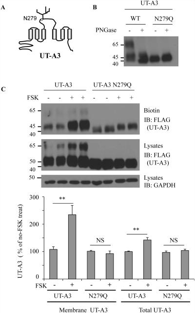
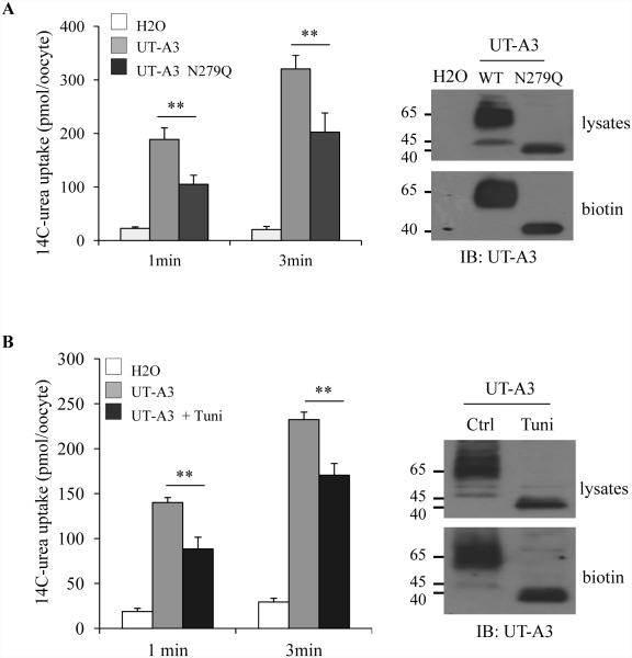
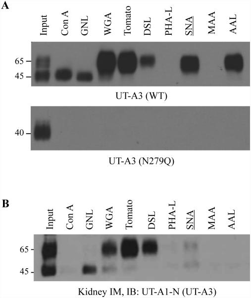
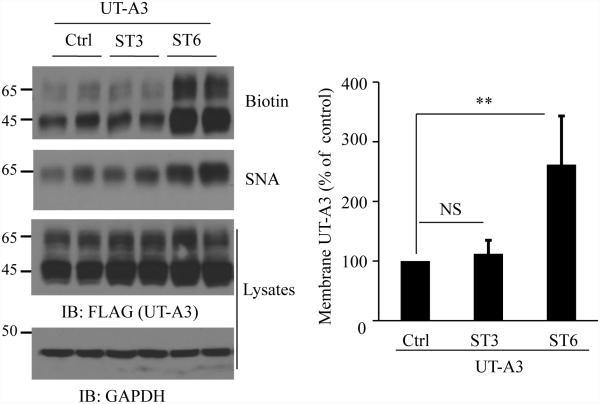
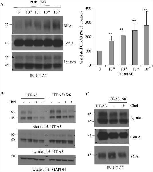
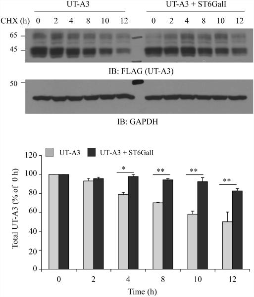
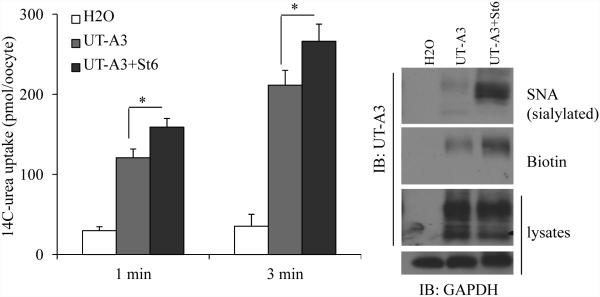
Two urea transporters, UT-A1 and UT-A3, are expressed in the kidney terminal inner medullary collecting duct (IMCD) and are important for the production of concentrated urine. UT-A1, as the largest isoform of all UT-A urea transporters, has gained much attention and been extensively studied; however, the role and the regulation of UT-A3 are less explored. In this study, we investigated UT-A3 regulation by glycosylation modification. A site-directed mutagenesis verified a single glycosylation site in UT-A3 at Asn279. Loss of the glycosylation reduced forskolin-stimulated UT-A3 cell membrane expression and urea transport activity. UT-A3 has two glycosylation forms, 45 and 65 kDa. Using sugar-specific binding lectins, the UT-A3 glycosylation profile was examined. The 45-kDa form was pulled down by lectin concanavalin A (Con A) and Galant husnivalis lectin (GNL), indicating an immature glycan with a high amount of mannose (Man), whereas the 65-kDa form is a mature glycan composed of acetylglucosamine (GlcNAc) and poly-N-acetyllactosame (poly-LacNAc) that was pulled down by wheat germ agglutinin (WGA) and tomato lectin, respectively. Interestingly, the mature form of UT-A3 glycan contains significant amounts of sialic acid. We explored the enzymes responsible for directing UT-A3 sialylation. Sialyltransferase ST6GalI, but not ST3GalIV, catabolizes UT-A3 α2,6-sialylation. Activation of protein kinase C (PKC) by PDB treatment promoted UT-A3 glycan sialylation and membrane surface expression. The PKC inhibitor chelerythrine blocks ST6GalI-induced UT-A3 sialylation. Increased sialylation by ST6GalI increased UT-A3 protein stability and urea transport activity. Collectively, our study reveals a novel mechanism of UT-A3 regulation by ST6GalI-mediated sialylation modification that may play an important role in kidney urea reabsorption and the urinary concentrating mechanism.

Keywords: Glycosylation; Protein kinase C; Sialyltransferase; Urea transporter.


Figures

Figure 1
Figure 1. A single glycosylation site at N279 in UT-A3
A: Structure diagram showing a glycosylation site at N279 located in the large extracellular loop. B: PNGase F digestion. FLAG-UT-A3 WT and FLAG-UT-A3 N279 were transiently transfected into HEK293 cells. 2 days later, the cell membrane lipid raft fractions were collected and de-glycosylated by PNGase F at 37°C for 2h. UT-A3 was analyzed by Western blot with FLAG antibody. C: UT-A3 membrane expression. HEK293 cells were transiently transfected with UT-A3 WT or N279Q for 48h. Cells were pre-treated with 10 μM forskolin (FSK) for 20 min and processed for cell surface biotinylation. Membrane UT-A3 proteins were precipitated by steptavidin beads followed by Western blot with FLAG antibody. The bar graphs show the percent increase in UT-A3 protein levels resulting from FSK treatment (** p<0.01, NS: no significance, n=3).
Figure 2
Figure 2. Un-glycosylated UT-A3 has reduced urea transport activity
Oocytes were injected with UT-A3 or UT-A3 N279Q cRNAs (A) or pre-injected with 10 ng tunicamycin (Tuni) then injected with UT-A3 cRNA (B). After 3 days. Urea transport activity was measured by 14C-labeled urea flux (n=6 oocyte/time point; mean ± SD). UT-A3 protein expression on the cell membrane was examined by biotinylation and followed by Western blot with a UT-A1 NH2-terminal antibody. A representative blot was shown from 3 different experiments.
Figure 3
Figure 3. Lectin affinity analysis of UT-A3 glycans
HEK293 cells transfected with UT-A3 WT or N279Q (A) or rat kidney inner medulla (IM) tissues (B) were processed for lipid raft isolation by 5%-40% (v/v) sucrose gradient ultracentrifugation. Lipid raft fractions (2-5) were collected for lectin pull-down assay and followed by Western blot with antibodies to UT-A1 NH2-terminus. A representative blot was shown from >3 different experiments.
Figure 4
Figure 4. Sialytransferase ST6GalI directs UT-A3 sialylation and cell membrane expression
UT-A3 membrane expression. HEK293 cells co-transfected UT-A3 without or with ST6GalI (ST6) or ST3GalIV (ST3) were processed for cell surface biotinylation. The total and the cell membrane UT-A3 proteins were examined by Western blot with FLAG antibody. UT-A3 protein sialylation was examined by SNA pull-down assay. Bar graph showed the cell membrane UT-A3 expression as a percent of control (** P<0.01, n=4).
Figure 5
Figure 5. Activation of PKC pathway promotes UT-A3 sialylation
A: UT-A3 sialylation by PDB. HEK 293 cells were transiently transfected with UT-A3 for 48h followed by PDBu treatment for 4h. Sialylated UT-A3 was pulled down by SNA and then examined by western blot. Con A lectin was used as a control. Bar graph showed the band densities of sialylated UT-A3 as a percent of control (0 PDBu) (** P<0.01, n=3). B: UT-A3 membrane expression. HEK293 cells were transiently transfected with UT-A3 alone or together with ST6GalI for 48h, and then treated with or without 2 μM chelerythrine (chel) for 2h. Cell surface proteins were biotinylated and analyzed by Western blot with FLAG antibody. C: Inhibition of ST6GalI-induced sialylation by PKC inhibitor. HEK293 cells transfected with UT-A3 alone or together with ST6GalI for 48h, then treated with chelerythrine for 2 h, sialylated UT-A3 was pulled down by SNA and then examined by western blot. B, C showed the representative western blots from three independent experiments.
Figure 6
Figure 6. Effect of ST6GalI on UT-A3 protein degradation
HEK293 cells transfected with UT-A3 alone or together with ST6GalI were treated with 100 μg/ml cycloheximide (CHX) for the indicated time. The cells were lysed with RIPA buffer and total UT-A3 protein levels were evaluated by Western blot with FLAG antibody. Bar graph showed the band densities of UT-A3 (including both 65- and 45-kDa) as a percent of control (0 h) (* P<0.05, ** P<0.01, n=3).
Figure 7
Figure 7. Effect of ST6GalI on UT-A3 activity
Oocytes were injected with cRNAs of UT-A3 (2 ng/cell) alone or plus ST6GalI (2 ng/cell). After 3 days, rea transport activity was measured by 14C-labeled urea flux (n=6 oocyte/time point; mean ± SD). UT-A3 protein expression on the cell membrane was examined by biotinylation and sialylated UT-A3 was pulled down by SNA, followed by Western blot with a UT-A1 NH2-terminal antibody (experiments n=3).

Similar articles

Cited by

References

    1. Alli AA, Bao HF, Liu BC, Yu L, Aldrugh S, Montgomery DS, Ma HP, Eaton DC. Calmodulin and CaMKII modulate ENaC activity by regulating the association of MARCKS and the cytoskeleton with the apical membrane. Am J Physiol Renal Physiol. 2015;309:F456–463. - PMC - PubMed
    1. Blessing NW, Blount MA, Sands JM, Martin CF, Klein JD. Urea transporters UT-A1 and UT-A3 accumulate in the plasma membrane in response to increased hypertonicity. Am J Physiol Renal Physiol. 2008;295:F1336–1341. - PMC - PubMed
    1. Blount MA, Klein JD, Martin CF, Tchapyjnikov D, Sands JM. Forskolin stimulates phosphorylation and membrane accumulation of UT-A3. Am J Physiol Renal Physiol. 2007;293:F1308–1313. - PubMed
    1. Bradford AD, Terris JM, Ecelbarger CA, Klein JD, Sands JM, Chou CL, Knepper MA. 97- and 117-kDa forms of collecting duct urea transporter UT-A1 are due to different states of glycosylation. Am J Physiol Renal Physiol. 2001;281:F133–143. - PubMed
    1. Breen KC, Georgopoulou N. The role of protein phosphorylation in alpha2,6(N)-sialyltransferase activity. Biochem Biophys Res Commun. 2003;309:32–35. - PubMed

Publication types

LinkOut - more resources