. 2016 Apr 5;24(4):518-527.
doi: 10.1016/j.str.2016.02.001. Epub 2016 Mar 10.

Unlocking the Bacterial SecY Translocon

Affiliations

Unlocking the Bacterial SecY Translocon

Robin A Corey et al. Structure. .

Abstract

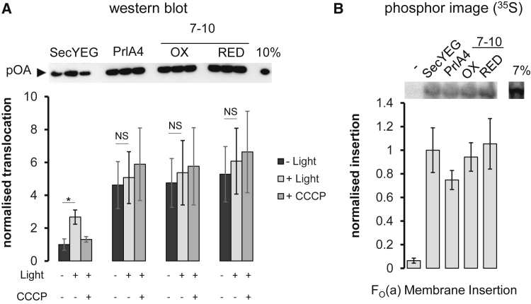
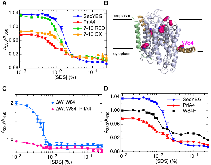
The Sec translocon performs protein secretion and membrane protein insertion at the plasma membrane of bacteria and archaea (SecYEG/β), and the endoplasmic reticular membrane of eukaryotes (Sec61). Despite numerous structures of the complex, the mechanism underlying translocation of pre-proteins, driven by the ATPase SecA in bacteria, remains unresolved. Here we present a series of biochemical and computational analyses exploring the consequences of signal sequence binding to SecYEG. The data demonstrate that a signal sequence-induced movement of transmembrane helix 7 unlocks the translocon and that this conformational change is communicated to the cytoplasmic faces of SecY and SecE, involved in SecA binding. Our findings progress the current understanding of the dynamic action of the translocon during the translocation initiation process. The results suggest that the converging effects of the signal sequence and SecA at the cytoplasmic face of SecYEG are decisive for the intercalation and translocation of pre-protein through the SecY channel.


Figures

None
Graphical abstract
Figure 1
Figure 1
Structure of the SecY Complex and Previously Proposed Effects of Signal Sequence Binding (A) Top left: M. jannaschii SecYEβ viewed from the outside (equivalent to the bacterial periplasm) (van den Berg et al., 2004). SecY TMs 1–5 are white; TMs 6–10 are blue; SecE is beige and Secβ is light green (equivalent to the E. coli SecG). In SecY, the plug (red) and TM 7 (turquoise) have been highlighted. Positions of SecY I284 and T404, which are crosslinked to form SecY7–10EG, are shown as orange and green circles, respectively. Top right: E. coli SecYEG bound to a signal sequence (SS; magenta) (Hizlan et al., 2012), coloring as in the equivalent view of the M. jannaschii SecYEβ (top left). The colored arrows highlight the substantial rearrangement of the plug and TM 7 upon SS binding. Bottom left: as per the top left panel but viewed from the side and with TMs 1–5 of SecY, SecE, and SecG removed for clarity. The approximate position of the membrane is marked by black lines. Bottom right: as per the bottom left panel but of the SS-bound structure, with the movement of the plug and TM 7 shown by colored arrows. The SS causes a distinctive tilting of TM 7. (B) Schematic diagram of SecY showing the ten TMs, along with the plug domain (labeled “p”) and loops C4 and C5. The position of the amino acid substitutions used in this study are shown as red circles for SecYprlA4EG, orange and green circles for SecY710EG, and green letters for the RPG motif. The trypsin cleavage site on SecY is indicated with a pair of scissors. See also Figure S1.
Figure 2
Figure 2
Competing Effects of the Activating Variant prlA4 and Inactivating Variant SecYEDPEG (A) SecYEG, SecYprlA4EG (PrlA4), SecYEDPEG (EDP), and SecYprlA4-EDPEG (PrlA4-EDP) were subjected to trypsin digestion followed by Coomassie-stained SDS-PAGE to examine the effect of different amino acid substitutions on the conformation of the C4 loop of SecY (the site of a prominent tryptic cleavage site). Samples are either untreated (−) or treated with 0.075 μg/ml (+) or 0.75 μl/ml (++) trypsin. The major bands correspond to full-length SecY, the N-terminal cleavage fragment, the C-terminal fragment, SecE, and a lower band for both SecE breakdown products and secondary SecY cleavage products (asterisk). (B) The rate of ATP turnover (kcat) is shown for SecA bound to different variants of SecYEG reconstituted into PL (purple), and following addition of a translocation substrate pOA (blue). ATP turnover is diminished by the C5 substitutions (SecYEDPEG), but is then restored when combined with the prlA4 mutations. Error bars denote SEM of three repeats. (C) Translocation of pOA into PL reconstituted with different SecYEG variants. Translocation efficiencies were determined by western blotting against pOA following a protease K protection assay (transported pOA is in the PL interior and protected from proteolysis); for a representative blot see Figure S3A. Results were quantified against a non-protease K-treated control and normalized to standard SecYEG, shown with SEM of three repeats. As in (B), the abolished activity of SecYEDPEG (EDP) is rescued by the prlA4 mutations. (D) Affinities of SecYEG for SecA in detergent solution, determined through quenching of a fluorescent marker on SecA (SecA). The decrease in fluorescence was plotted and fitted to tight binding equations (raw data are shown in Figure S3B). The calculated KD values are shown with SEM of three runs. ∗∗p = 0.004. (E) Representative structures of the cytoplasmic face of SecY (cartoon representation) following MD simulations of SecYEG, SecYEDPEG, and SecYprlA4-EDPEG. Structures are viewed from the side, with the cytosolic face indicated by the black line. The C4 loops are shown in light blue, the C5 loops green, and the rest of SecY white. In each panel, the position of the Cα of the conserved arginine in the RPG motif (mutated to glutamic acid in the middle and right-hand panels) is shown as a green sphere and labeled “R” or “E” accordingly. The approximate position of the trypsin cleavage sites is indicated by a pair of scissors. These structural changes are quantified in Figure S3D.
Figure 3
Figure 3
Comparative Secretion and Membrane Protein Insertion Activity of the SecYEG Variants (A) Post-translational translocation of the pre-protein substrate pOA into the interior of PL co-reconstituted with SecYEG and the light-driven proton pump bacteriorhodopsin (BR), with or without PMF (generated by bR: + Light or − Light). The transport efficiencies were determined by western blotting against pOA, and normalized to SecYEG without light; the SEM is shown for three repeats. p = 0.018; NS, not significant. (B) Co-translational membrane protein insertion of subunit a of the F1FO-ATP synthase (FO(a)) into the membranes of PL containing variants of SecYEG. The insertion levels were determined by the quantification of radiolabeled [35S]methionine incorporated into the newly synthesized and membrane inserted FO(a) substrate, averaged over three runs with SEM shown by the error bars.
Figure 4
Figure 4
Conformational Change of SecYEG Monitored by the Intrinsic Fluorescence of the Native Tryptophan Residues (A) Changes in tryptophan fluorescence emission were recorded upon SDS titration for different SecYEG variants in detergent solution. The y axis shows the ratio of the fluorescence emission at 330 and 350 nm after excitation at 288 nm. Error bars are the SEM of three repeats. (B) Positions of native tryptophan residues in SecYEG (carbon atoms as magenta spheres, nitrogen atoms as blue spheres), shown on an E. coli homology model based on the M. jannaschii SecYEβ structure (1RHZ; van den Berg et al., 2004). Note that one of the E. coli tryptophan residues on SecE is missing as this TM is not present in M. jannaschii. The position of W84 is marked. (C) Fluorescence unfolding profile of SecYEG as in (A), but with the single tryptophan variant (SecYW84EG; W84) and all other tryptophans changed to phenylalanine, in either standard SecYEG or prlA4 background. Error bars are the SEM of three repeats. (D) Fluorescence unfolding profile of SecYEG as in (A), but with W84 substituted with phenylalanine and all other tryptophan residues retained (black line). The data for the experiment with standard SecYEG and SecYprlA4EG are shown for comparison (the same data as in A). Error bars are the SEM of three repeats.
Figure 5
Figure 5
Molecular Dynamics Simulations of the SecY Complex (A) Post-simulation snapshots of the M. jannaschii SecYEβ variants with the whole complex shown in gray except the amphipathic helix of SecE, which is colored according to variant: standard SecYEβ in blue; PrlA4 in red; oxidized SecY7–10Eβ in orange and reduced SecY7–10Eβ in green. The immobile region of SecY used for distance analysis in (B) is highlighted with a yellow box. The equivalent position of W84 is marked with a cyan asterisk. (B) Distance analysis between the SecE amphipathic helix and a rigid region of SecY (yellow box in A). The first 20 ns of simulation are shown in detail in Figure S6A. The distance for the input structure is shown by the black dotted line. In the right panel, the single oxidized SecY7-10EG simulation, which resembles the reduced states, is marked with a light orange arrow. (C) Crystal structures of the SecY complex alone, M. jannaschii SecYEβ (1RHZ) (van den Berg et al., 2004), and SecY bound to SecA, T. maritima SecYEG-A (3DIN) (Zimmer et al., 2008) (SecA not shown). The amphipathic helix of SecE is shown in both structures, respectively in blue and red, and the equivalent key tryptophan residue of the E. coli SecY-W84 (W20 in T. maritima) is highlighted by cyan spheres.
Figure 6
Figure 6
Model of SecYEG Unlocking and Activation by the Cooperative Action of the Signal Sequence and SecA Resting: based on the closed structure, as seen in 1RHZ (van den Berg et al., 2004), with SecY in light blue, the lateral gate (LG) helices TM 2 and TM 7 in dark blue and green, respectively, and the plug helix in red. The amphipathic helix and TM 3 of SecE are shown in yellow at the back of SecY. Note that the model has not described the dissociation of SecA dimers known to occur upon the interaction with SecYEG (Or et al., 2002). SecA bound: based on the structure of SecYEG-A (3DIN) (Zimmer et al., 2008). Monomeric SecA (red) has bound to SecYEG, causing a widening of the LG and a tilting in the SecE amphipathic helix. In this state the SS of the pre-protein (magenta) is well positioned to associate at the binding site at the SecY LG. Unlocked: based on the SS-bound SecYEG structure (Hizlan et al., 2012) and the analyses described here. The SS has bound to the LG of SecY, and caused a straightening of TM 7 and a release of the SecY plug. SS binding results in conformational changes in the C4 and C5 loops and the SecE amphipathic helix that could favor the subsequent association with SecA, which is yet to fully engage. The coloured arrows represent the conformational changes described either here or in Hizlan et al. (2012), and are coloured as per the region of the complex that they relate to. Note that it is not clear in which order SecA and SS binding occur. Open: from either an unlocked or SecA-bound state the channel is then fully primed for ATP-driven protein translocation. The structure of this open state may be similar to the recent structure of the Sec61 complex engaged with a nascent pre-secretory substrate (Voorhees and Hegde, 2016), except that in this case the pre-protein is presented by SecA rather than the ribosome.

References

    1. Alami M., Dalal K., Lelj-Garolla B., Sligar S.G., Duong F. Nanodiscs unravel the interaction between the SecYEG channel and its cytosolic partner SecA. EMBO J. 2007;26:1995–2004. - PMC - PubMed
    1. Arkowitz R.A., Joly J.C., Wickner W. Translocation can drive the unfolding of a preprotein domain. EMBO J. 1993;12:243–253. - PMC - PubMed
    1. Auer J., Spicker G., Böck A. Presence of a gene in the archaebacterium Methanococcus vannielii homologous to secY of eubacteria. Biochimie. 1991;73:683–688. - PubMed
    1. Beck K., Wu L.F., Brunner J., Muller M. Discrimination between SRP- and SecA/SecB-dependent substrates involves selective recognition of nascent chains by SRP and trigger factor. EMBO J. 2000;19:134–143. - PMC - PubMed
    1. Berendsen H.J.C., van der Spoel D., van Drunen R. GROMACS: a message-passing parallel molecular dynamics implementation. Computer Phys. Commun. 1995;91:43–56.

Publication types

MeSH terms