. 2016 Oct 6;35(40):5237-5247.
doi: 10.1038/onc.2016.58. Epub 2016 Mar 14.

Olfactomedin 4 deletion induces colon adenocarcinoma in ApcMin/+ mice

Affiliations

Olfactomedin 4 deletion induces colon adenocarcinoma in ApcMin/+ mice

W Liu et al. Oncogene. .

Abstract

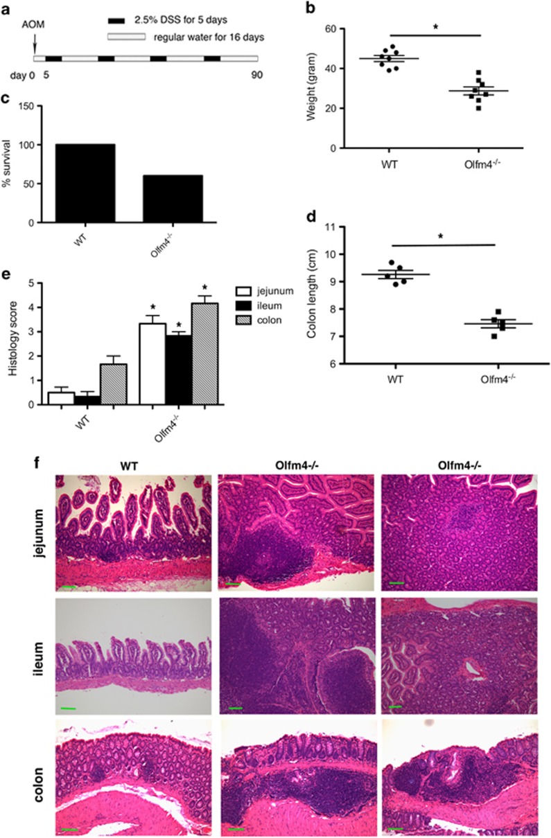
Colon carcinogenesis is a multiple-step process involving the accumulation of a series of genetic and epigenetic alterations. The most commonly initiating event of intestinal carcinogenesis is mutation of the adenomatous polyposis coli (APC) gene, which leads to activation of the Wnt/β-catenin pathway. Olfactomedin 4 (OLFM4) has emerged as an intestinal stem-cell marker, but its biological function in the intestine remains to be determined. Here we show that Olfm4 deletion induced colon adenocarcinoma in the distal colon of ApcMin/+ mice. Mechanistically, we found that OLFM4 is a target gene of the Wnt/β-catenin pathway and can downregulate β-catenin signaling by competing with Wnt ligands for binding to Frizzled receptors, as well as by inhibition of the Akt-GSK-3β (Akt-glycogen synthase kinase-3β) pathway. We have shown that both Wnt and nuclear factor-κB (NF-κB) signaling were boosted in tumor tissues of Apc Olfm4 double-mutant mice. These data establish OLFM4 as a critical negative regulator of the Wnt/β-catenin and NF-κB pathways that inhibits colon-cancer development initiated by APC mutation. In addition, Olfm4 deletion significantly enhanced intestinal-crypt proliferation and inflammation induced by azoxymethane/dextran sodium sulfate. Thus, OLFM4 has an important role in the regulation of intestinal inflammation and tumorigenesis, and could be a potential therapeutic target for intestinal malignant tumors. Unlike the human colonic epithelium, the mouse colonic epithelium does not express OLFM4, but nevertheless, systemic OLFM4 deletion promotes colon tumorigenesis and that loss from mucosal neutrophils may have a role to play.


Figures

Figure 1
Figure 1
Olfm4 deletion induces large polyps in the small intestine and adenocarcinoma formation in the distal colon of Apc+/− mice. (a) Polyp formation (arrow) in the small intestine of ApcMin/+, Apc+/− Olfm4+/− and Apc+/− Olfm4−/− (Apc Olfm4 double-mutant) mice. (b) Adenocarcinoma formation in the distal colon of Apc+/− Olfm4+/− and Apc Olfm4 double-mutant mice. Three representative tumors in mice of each genotype are presented. Hematoxylin and eosin (HE) histology staining is shown below each corresponding tumor. Scale bar, 50 μm. (c) Mean (±s.d.) small intestinal polyp number and size, and colon-tumor incidence in ApcMin/+ (n=20), Apc+/− Olfm4+/− (n=23) and Apc Olfm4 double-mutant mice (n=22) mice.
Figure 2
Figure 2
Olfm4 deletion enhances Wnt/β-catenin signaling in Apc Olfm4 double-mutant mice. (a–h) β-Catenin immunohistochemistry staining was performed in the distal colon of Apc+/+ Olfm4+/+ (WT) (a), Apc+/− Olfm4+/+ (b), Apc+/+ Olfm4−/− (c), Apc+/− Olfm4+/− (d) and Apc+/− Olfm4−/− (e and g) mice. Hematoxylin and eosin (HE) staining in (f and h) was performed in mice corresponding to (e and g), respectively. Scale bar, 50 μm. (i) A heatmap from cDNA microarray analysis shows Wnt pathway-related or target gene-expression level in the distal colon of mice with different genotypes. N, tumor-adjacent normal tissue; T, tumor. (j) Mean (±s.d.) (n=3) gene-expression levels for six representative Wnt target genes were confirmed by qRT–PCR. See also Supplementary Tables S1 and S2.
Figure 3
Figure 3
OLFM4 decreases β-catenin levels. (a–k) Western blots were performed in 293T cells (a–i), SW480 cells (j) and freshly isolated mouse colon crypts (k) after human OLFM4 treatment (1 μg/ml) (a–j) and mouse Olfm4 treatment (1 μg/ml) (k). β-Actin, total cadherin, Hsp90 and SP1 were used as loading controls in total cell lysate, membrane, cytoplasm and nucleus cell preparations, respectively. (a) β-Catenin levels in total cell lysate, membrane, cytoplasm and nucleus at different time points of OLFM4 treatment. (b) Endogenous and Wnt3a (1 μg/ml)-induced β-catenin levels in the cytoplasm and nucleus of cells pretreated with and without OLFM4 for 30 min. (c) Endogenous and Wnt3a (1 μg/ml)-induced β-catenin levels in the cytoplasm of cells transfected with control (vector) or OLFM4 expression plasmid. (d) β-Catenin phosphorylation at different time points of OLFM4 treatment. (e and f) Akt (T308 or Ser473) phosphorylation at different time points of OLFM4 treatment. (g and h) β-Catenin levels in the cytoplasm of cells pretreated with and without either LiCl (20 mm) or MG132 (10 μm) before OLFM4 treatment for 30 min. (i) β-Catenin levels in total cell lysates of cells transfected with Flag-tagged WT or mutant (S33Y or S31/37T41S45) β-catenin expression plasmids and then treated with or without OLFM4 for 30 min. (j) Total and active β-catenin levels in the cytoplasm or nucleus of SW480 colon-cancer cells at different time points of Olfm4 treatment. (k) β-Catenin levels in the cytoplasm and nucleus of freshly isolated mouse colon crypts at different time points of Olfm4 (or PBS control buffer) treatment. (l) Olfm4 and β-catenin levels in the cytoplasm of Ls174T cells infected with lentiviral control shRNA and Olfm4 shRNA.
Figure 4
Figure 4
OLFM4 interacts with Frizzled-7 and -10. (a) Ls174T cell lysate in the presence or absence of OLFM4 treatment was immunoprecipitated with OLFM4 antibody or IgG control antibody and then subjected to western blot with a total Frizzled (Fzd) receptors antibody. (b) Ls174T cell lysate was immunoprecipitated with OLFM4 antibody or IgG control antibody and then subjected to western blot with Fzd-7 or Fzd-10 antibody. (c) Ls174T cell lysate was immunoprecipitated with a total Fzd receptors antibody, Fzd-10 antibody or IgG antibody and then subjected to western blot with OLFM4 antibody. (d) Cell lysates from 293T cells stably overexpressing OLFM4 were immunoprecipitated with OLFM4 antibody or IgG control antibody and then subjected to western blot with a total Fzd receptors antibody, Fzd-7 antibody or Fzd-10 antibody. (e) Cell lysate from 293T cells stably overexpressing OLFM4 was immunoprecipitated with a total Fzd receptors antibody, Fzd-7 antibody, Fzd-10 antibody or IgG control antibody and then subjected to western blot with OLFM4 antibody. (f) Microscale thermophoresis (MST) measurements of the OLFM4 interaction with Fzd-7 (solid circles) and Fzd-10 (hollow circles). The solid lines are the best-fit curves to the data using the Hill equation (KD=2.5 μm for the OLFM4–Fzd-7 interaction and KD=10 μm for the OLFM4–Fzd-10 interaction). The error bars represent the s.d. from three independent measurements.
Figure 5
Figure 5
OLFM4 is a target gene of the Wnt/β-catenin/TCF pathway. (a) Three LEF1/TCF binding sites (in black box) located in the promoter of OLFM4. (b) The core (CAAAG or CTTTG) of the LEF1/TCF binding site was deleted individually or in combination as indicated by gray boxes. Data represent mean (±s.d.) (n=3) relative luciferase activity of corresponding reporter in the presence of Wnt3a or vehicle in 293T cells. *P<0.05 and **P<0.01 when compared with WT vehicle or WT Wnt3a, respectively, two-tailed t-test. (c) Mean (±s.d.) (n=3) OLFM4 mRNA expression determined by qRT–PCR in Ls174T cells (right) after knockdown of TCF4 expression by TCF4 lentivirus shRNA (left). *P<0.01 when compared with control (Con) shRNA, two-tailed t-test. (d) Mean (±s.d.) (n=3) OLFM4 mRNA expression determined by qRT–PCR in Ls174T and SW948 cells treated with IWP2, XAV-939 or DMSO control. *P<0.01 when compared with DMSO group, two-tailed t-test. (e) ChIP assay of LEF1 and TCF4 binding to all three LEF/TCF binding sites in the OLFM4 promoter.
Figure 6
Figure 6
Olfm4 deletion results in intestinal-crypt hyperplasia and severe inflammation after AOM/DSS treatment. (a) Schematic of AOM/DSS-induced colitis-associated cancer model. Mice were injected intraperitoneally with AOM (10 mg/kg). After 5 days, mice were treated with 2.5% DSS in their drinking water for 5 days followed by regular water for 16 days. The cycle was repeated four times. (b) WT (n=8) and Olfm4−/− (n=8) mice were weighed after four cycles of DSS and water treatment. *P<0.01, two-tailed t-test. (c) Percentage of survival of WT (n=10) and Olfm4−/− (n=10) mice after four cycles of DSS and water treatment. (d) Length of colon in WT (n=5) and Olfm4−/− (n=5) mice after four cycles of DSS and water treatment. *P<0.01, two-tailed t-test. (e) Mean (±s.d.) histology score of inflammation and tissue damage in the jejunum, ileum and colon of WT (n=8) and Olfm4−/− (n=8) mice after four cycles of DSS and water treatment. *P<0.01 when compared with WT corresponding tissues, two-tailed t-test. (f) HE staining of jejunum, ileum and colon of WT and Olfm4−/− mice after four cycles of DSS and water treatment. Scale bar, 50 μm. The image represents at least five mice in each group.

References

    1. Fearon ER. Molecular genetics of colorectal cancer. Annu Rev Pathol 2011; 6: 479–507. - PubMed
    1. Bienz M, Clevers H. Linking colorectal cancer to Wnt signaling. Cell 2000; 103: 311–320. - PubMed
    1. Inomata M, Ochiai A, Akimoto S, Kitano S, Hirohashi S. Alteration of beta- catenin expression in colonic epithelial cells of familial adenomatous polyposis patients. Cancer Res 1996; 56: 2213–2217. - PubMed
    1. Karin M, Greten FR. NF-kappaB: linking inflammation and immunity to cancer development and progression. Nat Rev Immunol 2005; 5: 749–759. - PubMed
    1. Schwitalla S, Fingerle AA, Cammareri P, Nebelsiek T, Goktuna SI, Ziegler PK et al. Intestinal tumorigenesis initiated by dedifferentiation and acquisition of stem-cell-like properties. Cell 2013; 152: 25–38. - PubMed

Publication types