

Effect of artesunate supplementation on bacterial translocation and dysbiosis of gut microbiota in rats with liver cirrhosis
- PMID: 26973391
- PMCID: PMC4779918
- DOI: 10.3748/wjg.v22.i10.2949
Effect of artesunate supplementation on bacterial translocation and dysbiosis of gut microbiota in rats with liver cirrhosis
Abstract
Aim: To evaluate the effect of artesunate (AS) supplementation on bacterial translocation (BT) and gut microbiota in a rat model of liver cirrhosis.
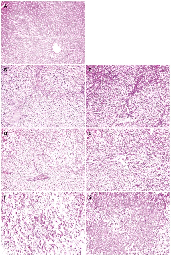
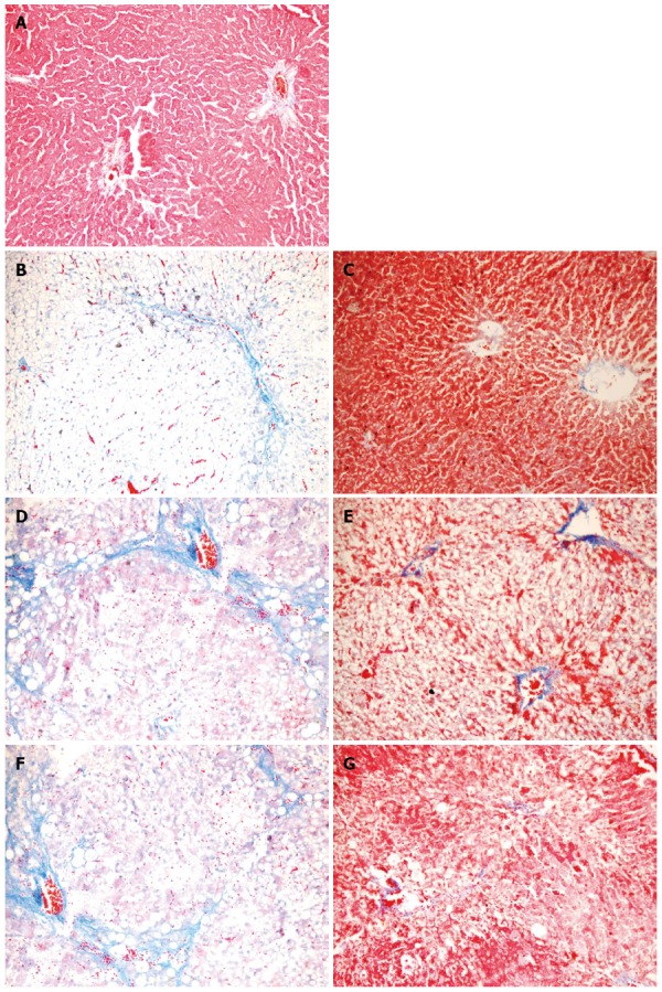
Methods: Fifty-four male Sprague-Dawley rats were randomly divided into a normal control group (N), a liver cirrhosis group (M) and a liver cirrhosis group intervened with AS (MA). Each group was sampled at 4, 6 and 8 wk. Liver cirrhosis was induced by injection of carbon tetrachloride (CCl4), intragastric administration of 10% ethanol, and feeding a high fat diet. Rats in the MA group were intragastrically administered with AS (25 mg/kg body weight, once daily). Injuries of the liver and intestinal mucosa were assessed by hematoxylin-eosin or Masson's trichrome staining. Liver index was calculated as a ratio of the organ weight (g) to body weight (g). The gut microbiota was examined by automated ribosomal intergenic-spacer analysis of fecal DNA. BT was assessed by standard microbiological techniques in the blood, mesenteric lymph nodes (MLNs), liver, spleen, and kidney.
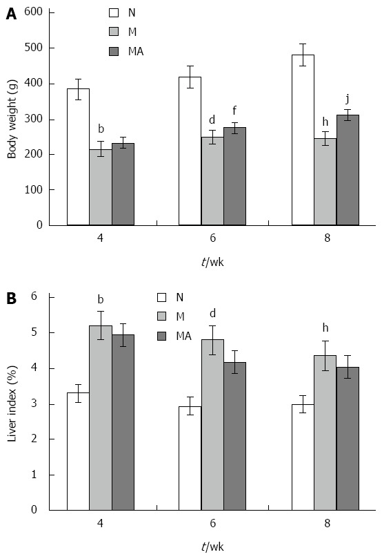
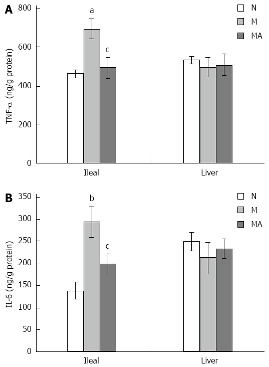
Results: Compared to group N, the body weight was reduced significantly in groups M and MA due to the development of liver cirrhosis over the period of 8 wk. The body weight was higher in group MA than in group M. The liver indices were significantly elevated at 4, 6 and 8 wk in groups M and MA compared to group N. AS supplementation partially decreased the liver indices in group MA. Marked histopathologic changes in the liver and small intestinal mucosa in group M were observed, which were alleviated in group MA. Levels of pro-inflammatory interleukin-6 and tumor necrosis factor-α were significantly elevated at 8 wk in ileal homogenates in group M compared to group N, which were decreased after AS supplementation in group MA. The dysbiosis of gut microbiota indicated by the mean diversity (Shannon index) and mean similarity (Sorenson index) was severe as the liver cirrhosis developed, and AS supplementation had an apparent intervention effect on the dysbiosis of gut microbiota at 4 wk. The occurrence of BT was increased in the liver of group M compared to that of group N. AS supplementation reduced BT in group MA at 8 wk. BT also occurred in the MLNs, spleen, and kidney, which was reduced by AS supplementation. BT was not detected in the blood in any group.
Conclusion: Dysbiosis of gut microbiota, injury of intestinal mucosal barrier and BT occurred as liver cirrhosis progressed, which might enhance inflammation and aggravate liver injury. AS may have other non-antimalarial effects that modulate gut microbiota, inhibit BT and alleviate inflammation, resulting in a reduction in CCl4, alcohol and high fat-caused damages to the liver and intestine.
Keywords: Artesunate; Bacterial translocation; Gut microbiota; Hepatic cirrhosis; Intervention.
Figures

Similar articles
-
Mosapride Stabilizes Intestinal Microbiota to Reduce Bacterial Translocation and Endotoxemia in CCl4-Induced Cirrhotic Rats.Dig Dis Sci. 2017 Oct;62(10):2801-2811. doi: 10.1007/s10620-017-4704-x. Epub 2017 Aug 16. Dig Dis Sci. 2017. PMID: 28815345
-
Norfloxacin is more effective than Rifaximin in avoiding bacterial translocation in an animal model of cirrhosis.Liver Int. 2018 Feb;38(2):295-302. doi: 10.1111/liv.13551. Epub 2017 Sep 13. Liver Int. 2018. PMID: 28834270
-
Obeticholic acid reduces bacterial translocation and inhibits intestinal inflammation in cirrhotic rats.J Hepatol. 2016 May;64(5):1049-1057. doi: 10.1016/j.jhep.2015.12.010. Epub 2015 Dec 23. J Hepatol. 2016. PMID: 26723896
-
Gut microbiota and host metabolism in liver cirrhosis.World J Gastroenterol. 2015 Nov 7;21(41):11597-608. doi: 10.3748/wjg.v21.i41.11597. World J Gastroenterol. 2015. PMID: 26556989 Free PMC article. Review.
-
Bacterial translocation in patients with liver cirrhosis: physiology, clinical consequences, and practical implications.Expert Rev Gastroenterol Hepatol. 2018 Jul;12(7):641-656. doi: 10.1080/17474124.2018.1481747. Epub 2018 Jun 6. Expert Rev Gastroenterol Hepatol. 2018. PMID: 29806487 Review.
Cited by
-
Vitamin D Improves Intestinal Barrier Function in Cirrhosis Rats by Upregulating Heme Oxygenase-1 Expression.Biomol Ther (Seoul). 2019 Mar 1;27(2):222-230. doi: 10.4062/biomolther.2018.052. Biomol Ther (Seoul). 2019. PMID: 30173501 Free PMC article.
-
Gut microbes combined with metabolomics reveal the protective effects of Qijia Rougan decoction against CCl4-induced hepatic fibrosis.Front Pharmacol. 2024 Mar 28;15:1347120. doi: 10.3389/fphar.2024.1347120. eCollection 2024. Front Pharmacol. 2024. PMID: 38606180 Free PMC article.
-
Resveratrol ameliorates liver fibrosis induced by nonpathogenic Staphylococcus in BALB/c mice through inhibiting its growth.Mol Med. 2022 May 4;28(1):52. doi: 10.1186/s10020-022-00463-y. Mol Med. 2022. PMID: 35508992 Free PMC article.
-
Efficacy of Biejiajian Pill on Intestinal Microbiota in Patients with Hepatitis B Cirrhosis/Liver Fibrosis: A Randomized Double-Blind Controlled Trial.Chin J Integr Med. 2023 Sep;29(9):771-781. doi: 10.1007/s11655-023-3542-2. Epub 2023 May 24. Chin J Integr Med. 2023. PMID: 37222832 Clinical Trial.
-
Protective Effects of Ethanolic Extracts from Artichoke, an Edible Herbal Medicine, against Acute Alcohol-Induced Liver Injury in Mice.Nutrients. 2017 Sep 11;9(9):1000. doi: 10.3390/nu9091000. Nutrients. 2017. PMID: 28891983 Free PMC article.
References
-
- Izcue A, Coombes JL, Powrie F. Regulatory lymphocytes and intestinal inflammation. Annu Rev Immunol. 2009;27:313–338. - PubMed
-
- MacDonald TT, Monteleone I, Fantini MC, Monteleone G. Regulation of homeostasis and inflammation in the intestine. Gastroenterology. 2011;140:1768–1775. - PubMed
-
- Lawrance IC. Microbiota and management of inflammatory bowel disease. J Gastroenterol Hepatol. 2012;27:1137–1140. - PubMed
-
- Fujimoto T, Imaeda H, Takahashi K, Kasumi E, Bamba S, Fujiyama Y, Andoh A. Decreased abundance of Faecalibacterium prausnitzii in the gut microbiota of Crohn’s disease. J Gastroenterol Hepatol. 2013;28:613–619. - PubMed
Publication types
MeSH terms
Substances
Grants and funding
LinkOut - more resources
Full Text Sources
Other Literature Sources
Research Materials
