. 2016 Mar 2:7:257.
doi: 10.3389/fpls.2016.00257. eCollection 2016.

Reconstructing the Origin of Oxygenic Photosynthesis: Do Assembly and Photoactivation Recapitulate Evolution?

Affiliations

Reconstructing the Origin of Oxygenic Photosynthesis: Do Assembly and Photoactivation Recapitulate Evolution?

Tanai Cardona. Front Plant Sci. .

Abstract

Due to the great abundance of genomes and protein structures that today span a broad diversity of organisms, now more than ever before, it is possible to reconstruct the molecular evolution of protein complexes at an incredible level of detail. Here, I recount the story of oxygenic photosynthesis or how an ancestral reaction center was transformed into a sophisticated photochemical machine capable of water oxidation. First, I review the evolution of all reaction center proteins in order to highlight that Photosystem II and Photosystem I, today only found in the phylum Cyanobacteria, branched out very early in the history of photosynthesis. Therefore, it is very unlikely that they were acquired via horizontal gene transfer from any of the described phyla of anoxygenic phototrophic bacteria. Second, I present a new evolutionary scenario for the origin of the CP43 and CP47 antenna of Photosystem II. I suggest that the antenna proteins originated from the remodeling of an entire Type I reaction center protein and not from the partial gene duplication of a Type I reaction center gene. Third, I highlight how Photosystem II and Photosystem I reaction center proteins interact with small peripheral subunits in remarkably similar patterns and hypothesize that some of this complexity may be traced back to the most ancestral reaction center. Fourth, I outline the sequence of events that led to the origin of the Mn4CaO5 cluster and show that the most ancestral Type II reaction center had some of the basic structural components that would become essential in the coordination of the water-oxidizing complex. Finally, I collect all these ideas, starting at the origin of the first reaction center proteins and ending with the emergence of the water-oxidizing cluster, to hypothesize that the complex and well-organized process of assembly and photoactivation of Photosystem II recapitulate evolutionary transitions in the path to oxygenic photosynthesis.

Keywords: anoxygenic photosynthesis; oxygenic photosynthesis; photoactivation; photoassembly; photosystem; reaction center; water oxidation.


Figures

Figure 1
Figure 1
Evolutionary relations of reaction center proteins. This scheme is based on the well-known phylogenetic relationships of reaction center proteins, which I have reviewed in detail before (Cardona, 2015). At the bottom right, the spheres with question marks represent the earliest evolutionary events that led to the evolution of the chlorophyll and bacteriochlorophyll synthesis pathway and the first reaction center proteins. It has been suggested that reaction center proteins might have originated from single-helix pigment-binding proteins (Allen and Vermaas, 2010) or from proteins related to the respiratory Cytochrome b proteins (Xiong and Bauer, 2002). Both hypotheses merit further consideration. The ancestral reaction center protein A gave rise to two new classes of proteins ancestral to Type I (I1) and II (II1), respectively. It is highly likely that these early stages of reaction center evolution occurred at a time well before the radiation of modern bacterial forms. Therefore, it is difficult to envision the mechanisms by which the ancestral reaction center evolved into two new forms and the evolutionary forces that aided such divergence. The subscript indicates transitional stages away from ancestral protein A. Both I1 and II1 separated into two new classes of proteins, one lineage led to the evolution of Photosystem I and II, employed in oxygenic photosynthesis and today only found in the phylum Cyanobacteria and photosynthetic eukaryotes. The other lineage led to the type of reaction center proteins employed by anoxygenic phototrophic bacteria. Type I reaction centers (longer rectangles) are characterized by having 11 transmembrane helices: the first 6 helices are the antenna domain in charge of light harvesting and the last 5 helices are the reaction center domain in charge of photochemistry. In Type II reaction centers (smaller rectangles) the first 6 helices are missing, and only the reaction center domain is found, the last 5 helices. Photosystem II is unique because it is associated with antenna proteins, the CP43 and CP47 subunits, which originated from a Type I reaction center. The nature of the ancestral reaction center protein A is uncertain, was it more like a Type I or Type II reaction center? Did it have an iron-sulfur cluster or a non-heme iron? Did it have an antenna domain or was this fused later with a reaction center protein to make the first Type I reaction centers? At the moment, from the existing structural and sequence data, it is not possible to answer these questions with certainty. Reaction center A probably had some traits from each reaction center type and also some unique traits no longer present in reaction center proteins today.
Figure 2
Figure 2
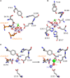
The Mn4CaO5 cluster of Photosystem II as resolved in the crystal structure by Umena et al. (2011), PDB ID: 3WU2. Panel (A) shows the cluster coordinated by the inner ligands D170 and E189 and the ligands provided by the CP43 subunit, E354, and R357. Panel (B) shows the ligands provided from the C-terminus of the D1 protein. Panel (C) shows a proposal for the high-affinity Mn binding site based on evolutionary grounds and supported by mutagenesis and spectroscopy (see text). After oxidation of the first Mn(II) to Mn(III), which might occur concomitantly with the deprotonation of a ligating water molecule, Ca2+ binds. The binding of Ca2+ shifts the initially bound Mn(III) to a position similar to that of Mn4 in the intact cluster.
Figure 3
Figure 3
Comparison of the antenna domain of Photosystem I reaction center proteins and the antenna proteins of Photosystem II from the crystal structures PDB ID: 1JB0 and 3WU2 from Thermosynechococcus spp., respectively. (A,B) highlight the core antenna of Photosystem I and II respectively (orange). Blue spheres highlight the conserved peripheral chlorophylls that allow excitation energy transfer from the core antenna to the reaction center, named ChlZ and ChlD in Photosystem II. (C–E) highlight the differences between the antenna domain of the PsaA subunit of Photosystem I with that of the CP43 and CP47, respectively. Note that the extrinsic domain is absent in PsaA. Panels (F,G) show and schematic representation of a Type I reaction center and a Photosystem II antenna protein. The alpha-helices have been color coded to show the homologous positions. Panels (H,I) show detail of the folding pattern of the extrinsic domain from the CP47 subunit. Panel (J) shows a sequence alignment of all Type I reaction center proteins including the CP47 and CP43 subunits. Only three fragments are shown for clarity, those corresponding to transmembrane helices E, F, and J in Type I reaction centers and their respective matching sequences in the antenna proteins of Photosystem II. The sequence alignment was performed using Clustal Omega that applies a Hidden Markov Model-based algorithm (Sievers et al., 2011). The full alignment is available on request.
Figure 4
Figure 4
Interaction of reaction center proteins and the small peripheral subunits. Panels (A–F) show the reaction center proteins of the anoxygenic Type II reaction center of Blastochloris viridis (M and L, PDB ID: 2PRC), Thermosynechococcus vulcanus (D1 and D2, PDB ID: 3WU2), and Thermosynechococcus elongatus (PsaA and PsaB, core domain only, PDB ID: 1JB0). The orange colored cartoon highlights the protein segment found between the 1st and 2nd transmembrane helices of the core proteins (7th and 8th in Photosystem I). Panels (G,H) show the position of the peripheral subunits of Photosystem II and Photosystem I. Panels (I–L) show the interaction of the core proteins with some of the small subunits and nearby pigments.
Figure 5
Figure 5
Comparison of the electron donor sides of Type II reaction centers from Thermosynechococcus vulcanus (PDB ID: 3WU2), with those from Blastochloris viridis (PDB ID: 1JB0), and Chloroflexus aggregans. The highlighted residues are at strictly homologous positions as determined by sequence and structural comparisons; the variation in number is due to the fact that different reaction center proteins are of different sizes in different strains of bacteria. The L and M from Chloroflexus aggregans are homology models made with the SWISS-MODEL service, automated mode, using the structure of the Blastochloris viridis reaction center as a template.
Figure 6
Figure 6
Sequence identity of Photosystem II reaction center proteins as a function of time. The blue dots show the percentage of sequence identity between the D1 protein sequence of Arabidopsis thaliana compared to that in other organisms. The red dots show the percentage of sequence identity of D2. The green blurred box shows the level of sequence identity between D1 and D2 of Arabidopsis and the blur represents uncertainty. The orange box shows the sequence identity of D2 compared to the M subunit of Chloroflexus aurantiacus. The gray box shows the sequence identity of D2 compared to the PsaB subunit of Photosystem I from Thermosynechococcus elongatus. This was calculated by overlaying the 3D structures of D2 and PsaB proteins and counting the conserved residues (the identity is below 5%), see Cardona (2015). The plot shows that D1 and D2 have been changing at an almost constant rate since the Great Oxygenation Event (GOE) around 2.4 billion years ago. It also highlights that the events that led to the divergence of Type I from Type II reaction centers (gray box), the anoxygenic from oxygenic Type II reaction centers (orange box), and D1 from D2 (green box), must have occurred very early in the history of life. It is also likely that these early events in the evolution of photosynthesis probably occurred relatively fast after the origin of the first reaction center protein. The divergence times in plant evolution were taken from Clarke et al. (2011). The time for Cyanophora paradoxa, representing the origin of plastids, was assumed to be in between 1.1 and 1.5 Ga (Butterfield, ; Yoon et al., 2004). The time for Nostoc sp. PCC 7120, representing the origin of heterocystous Cyanobacteria, was assumed to have occurred after the GOE (Golubic et al., ; Tomitani et al., 2006). The divergence of the genus Gloeobacter violaceus sp. PCC 7421 was assumed to have occurred before the GOE. The divergence time for D1/D2, D2/M, and D2/PsaB are hypothetical, but in order to explain the origin and diversification of photosynthesis within the age constraints of planet Earth, very fast rates of evolution are needed at the earliest stages. It suggests that the earliest stages of Photosystem II evolution, such as the divergence of D1 and D2, might have occurred soon after the origin of photochemical reaction centers.

References

    1. Allen J. F., Vermaas W. (2010). Evolution of photosynthesis, in Encyclopedia of Life Sciences (els), (Chichester: John Wiley & Sons, Ltd; ). Available online at: http://jfallen.org/publications/pdf/Allen_2010_ELS.pdf
    1. Allen J. F. (2005). A redox switch hypothesis for the origin of two light reactions in photosynthesis. FEBS Lett. 579, 963–968. 10.1016/j.febslet.2005.01.015 - DOI - PubMed
    1. Ananyev G., Nguyen T., Putnam-Evans C., Dismukes G. C. (2005). Mutagenesis of CP43-arginine-357 to serine reveals new evidence for (bi)carbonate functioning in the water-oxidizing complex of Photosystem II. Photochem. Photobiol. Sci. 4, 991–998. 10.1039/b507519j - DOI - PubMed
    1. Asada M., Mino H. (2015). Location of the high-affinity Mn2+ site in Photosystem II detected by peldor. J. Phys. Chem. B 119, 10139–10144. 10.1021/acs.jpcb.5b03994 - DOI - PubMed
    1. Baymann F., Brugna M., Mühlenhoff U., Nitschke W. (2001). Daddy, where did (PS)I come from? Biochim. Biophys. Acta 1507, 291–310. 10.1016/S0005-2728(01)00209-2 - DOI - PubMed