

Case Reports
doi: 10.1016/j.radcr.2015.12.004.
eCollection 2016 Mar.
Sonographic and magnetic resonance imaging findings of neurocutaneous melanosis
Affiliations
- PMID: 26973729
- PMCID: PMC4769615
- DOI: 10.1016/j.radcr.2015.12.004
Item in Clipboard
Case Reports
Sonographic and magnetic resonance imaging findings of neurocutaneous melanosis
Radiol Case Rep.
.
Abstract
Neurocutaneous melanosis is a rare nonfamilial phakomatosis characterized by large or multiple congenital melanocytic nevi plus the presence of central nervous system melanosis or melanoma. We report a case of a male infant with a giant posteroaxial nevus and evidence of intracranial melanosis on ultrasound and magnetic resonance imaging.
Keywords: Congenital nevus; MRI; Neonatal brain ultrasound; Neurocutaneous melanosis.
Figures
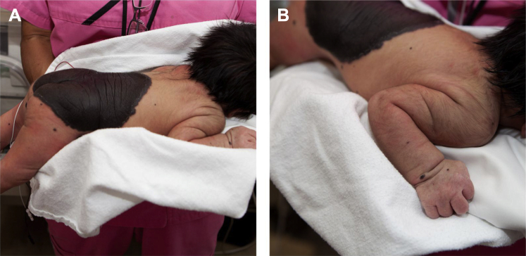
Congenital giant nevi on the back of a term baby, with dominant “cape-like” lesion on his back and buttocks (A), and multiple surrounding small lesions extending to both upper and lower extremities (B).
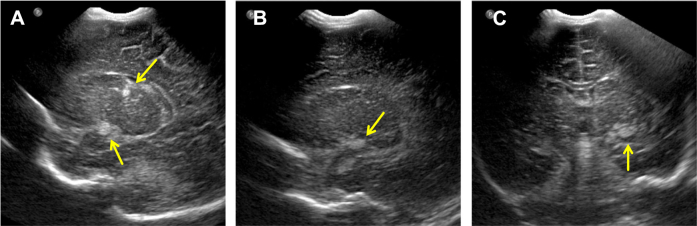
Neonatal transfontanellar ultrasound images in left parasagittal (A, B) and coronal (C) planes demonstrate echogenic lesions in the left thalamus and left choroidal fissure (arrows).
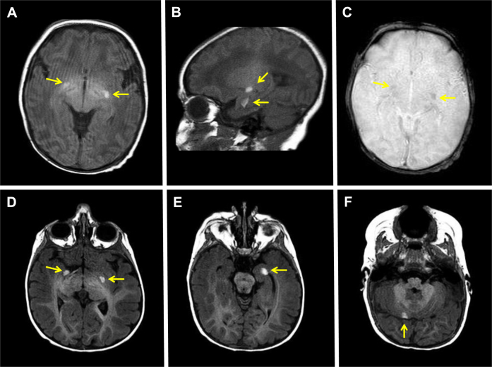
Neonatal brain MR images show multiple T1-hyperintense intraparenchymal lesions in bilateral inferior basal ganglia (A), left thalamus and left mesial temporal lobe (B). The lesions are mildly hypointense on susceptibility-weighted images (C). Repeat MR images, 3 months later, demonstrate stable T1-hyperintense lesions in bilateral inferior basal ganglia (D), left mesial temporal lobe (E), and right cerebellum (F). Arrows point to the intraparenchymal lesions.
References
-
- Rokitansky J. An excellent case of a pigmented mole with effused pigmentation of the inner meninges of the brain and the spinal cord. Allg Wien Mediz. 1861;26:113–116. [in German]
-
- Kadonaga J.N., Frieden I.J. Neurocutaneous melanosis: definition and review of the literature. J Am Acad Dermatol. 1991;24(5 Pt 1):747–755. - PubMed
-
- DeDavid M., Orlow S.J., Provost N., Marghoob A.A., Rao B.K., Huang C.L. A study of large congenital melanocytic nevi and associated malignant melanomas: review of cases in the New York University Registry and the world literature. J Am Acad Dermatol. 1997;36(3 Pt 1):409–416. - PubMed
-
- DeDavid M., Orlow S.J., Provost N., Marghoob A.A., Rao B.K., Wasti Q. Neurocutaneous melanosis: clinical features of large congenital melanocytic nevi in patients with manifest central nervous system melanosis. J Am Acad Dermatol. 1996;35(4):529–538. - PubMed
-
- Marghoob A.A., Dusza S., Oliveria S., Halpern A.C. Number of satellite nevi as a correlate for neurocutaneous melanocytosis in patients with large congenital melanocytic nevi. Arch Dermatol. 2004;140(2):171–175. - PubMed
Publication types
LinkOut - more resources
Full Text Sources
Other Literature Sources
