

Childhood giant omental and mesenteric lipoma
- PMID: 26973731
- PMCID: PMC4769613
- DOI: 10.1016/j.radcr.2015.12.003
Childhood giant omental and mesenteric lipoma
Abstract
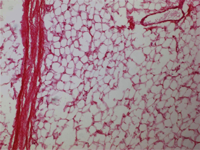
Omental and mesenteric lipomas are very rare benign lesions of mature adipose tissue. They are well-defined, noninvasive, and encapsulated masses that can be discovered in asymptomatic patients or may cause variable nonspecific symptoms depending on their size and location. The omental and mesenteric lipoma has confusing features in ultrasound; however, computed tomography and magnetic resonance imaging can well characterize and demarcate these lesions. Though few cases of mesenteric and omental lipomas have been reported in the literature, but because of its large size and childhood presentation, the case we present, can be one of the largest childhood omental and mesenteric lipomas ever reported. A 6-year-old girl presented with slowly progressing abdominal distension and repeated dull abdominal pain for last 4 years. Abdominal and pelvic computed tomography examination revealed a huge mesenteric and omental lipoma that was resected surgically without any complications.
Keywords: Abdominal distension; Lipoma; Mesenteric and omental mass; Pediatric abdominal mass.
Figures


References
-
- Sirikci A., Bayram M., Kervancioglu R., Sarica K. Abdominopelvic lipomatosis in a child with indefinite physical findings. Pediatr Radiol. 2000;30(7):480. - PubMed
-
- Rhydholm A., Berg N.O. Size, site and clinical incidence of lipoma. Acta Orthop Scand. 1983;54:929–934. - PubMed
-
- Prando A., Wallace S., Marins J.L., Pereira R.M., de Oliveira E.R., Alvarenga M. Sonographic features of benign intraperitoneal lipomatous tumor in children: report of 4 cases. Pediatr Radiol. 1990;20:571–574. - PubMed
Publication types
LinkOut - more resources
Full Text Sources
Other Literature Sources
