Retrospective Review of Ocular Point-of-Care Ultrasound for Detection of Retinal Detachment
- PMID: 26973752
- PMCID: PMC4786246
- DOI: 10.5811/westjem.2015.12.28711
Retrospective Review of Ocular Point-of-Care Ultrasound for Detection of Retinal Detachment
Abstract
Introduction: Retinal detachment is an ocular emergency that commonly presents to the emergency department (ED). Ophthalmologists are able to accurately make this diagnosis with a dilated fundoscopic exam, scleral depression or ophthalmic ultrasound when a view to the retina is obstructed. Emergency physicians (EPs) are not trained to examine the peripheral retina, and thus ophthalmic ultrasound can be used to aid in diagnosis. We assessed the accuracy of ocular point-of-care ultrasound (POCUS) in diagnosing retinal detachment.
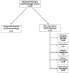
Methods: We retrospectively reviewed charts of ED patients with suspected retinal detachment who underwent ocular POCUS between July 2012 and May 2015. Charts were reviewed for patients presenting to the ED with ocular complaints and clinical concern for retinal detachment. We compared ocular POCUS performed by EPs against the criterion reference of the consulting ophthalmologist's diagnosis.
Results: We enrolled a total of 109 patients. Of the 34 patients diagnosed with retinal detachment by the ophthalmologists, 31 were correctly identified as having retinal detachment by the EP using ocular POCUS. Of the 75 patients who did not have retinal detachment, 72 were ruled out by ocular POCUS by the EP. This resulted in a POCUS sensitivity of 91% (95% CI [76-98]) and specificity of 96% (95% CI [89-99]).
Conclusion: This retrospective study suggests that ocular POCUS performed by EPs can aid in the diagnosis of retinal detachment in ED.
Figures
References
-
- Walker RA, Adhikari S. Eye Emergencies. Tintinalli’s Emergency Medicine: A Comprehensive Study Guide. 2011;236:1517–49.
-
- Alotaibi AG, Osman EA, Allam KH, et al. One month outcome of ocular related emergencies in a tertiary hospital in central Saudi Arabia. Saudi Med J. 2011;32:1256–60. - PubMed
-
- Mitry D, Charteris DG, Fleck BW, et al. The epidemiology of rhegmatogenous retinal detachment: geographical variation and clinical associations. Br J Ophthalmol. 2009;94(6):678–84. - PubMed
-
- Haimann MH, Burton TC, Brown CK. Epidemiology of retinal detachment. Arch Ophthalmol. 1982;100:289–92. - PubMed
Publication types
MeSH terms
LinkOut - more resources
Full Text Sources
Other Literature Sources
Medical


