

Evaluation of proliferation and cytokines production by mitogen-stimulated bovine peripheral blood mononuclear cells
- PMID: 26973760
- PMCID: PMC4769330
Evaluation of proliferation and cytokines production by mitogen-stimulated bovine peripheral blood mononuclear cells
Abstract
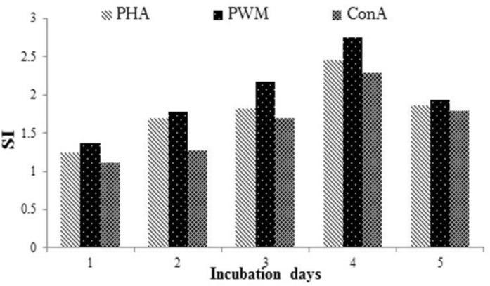
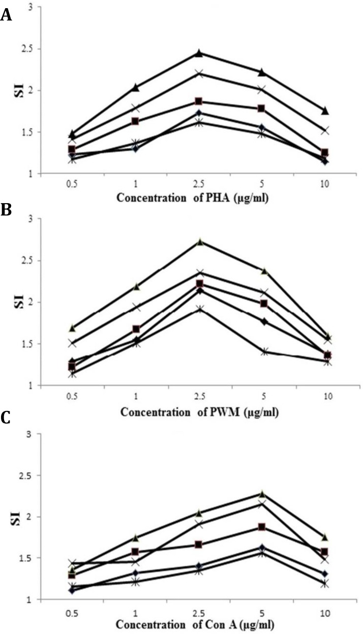
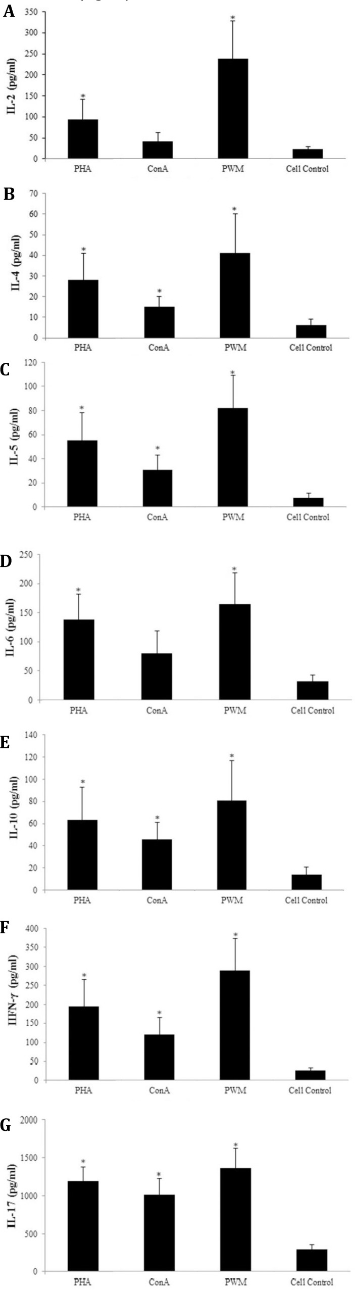
This in vitro study was conducted to evaluate lymphocyte blastogenic and cytokine production by bovine peripheral blood mononuclear cells (PBMCs) stimulated with phytohemagglutinin (PHA), pokeweed mitogen (PWM) and concanavalin A (Con A) mitogens, by using tetrazolium salt and ELISA tests, respectively. The results presented that Interleukin-2 (IL-2), IL-4, IL-5, IL-10, IL-17 and IFN-γ production in response to PWM mitogens was the highest and Con A the lowest amount and the median values of three mitogens were in the following order: PWM > PHA > Con A > cell control. In the case of IL-6, the production of this cytokine was the same amount for PWM and Con A and a lower amount for PHA stimulation. The results of this study not only showed a normal range for the production of these cytokines from PBMCs that were affected by mitogens, but it demonstrated that the bovine immune system at 2.5 to 3 months was post-natally matured enough to mount an effective immune response to mitogens as well as specific antigens.
Keywords: Bovine; Concanavalin A; Cytokine; Phytohemagglutinin; Pokeweed mitogen.
Figures
Similar articles
-
Mitogen-Induced Interferon Gamma Production in Human Whole Blood: The Effect of Heat and Cations.Curr Pharm Biotechnol. 2019;20(7):562-572. doi: 10.2174/1389201020666190528093432. Curr Pharm Biotechnol. 2019. PMID: 31132974
-
Antithrombin III inhibits lymphocyte proliferation, immunoglobulin production and mRNA expression of lymphocyte growth factors (IL-2, gamma-IFN and IL-4) in vitro.Transpl Immunol. 2001 Oct;9(1):1-6. doi: 10.1016/s0966-3274(01)00042-9. Transpl Immunol. 2001. PMID: 11680566
-
Development of a lymphocyte-transformation-assay for peripheral blood lymphocytes of the harbor porpoise and detection of cytokines using the reverse-transcription polymerase chain reaction.Vet Immunol Immunopathol. 2004 Mar;98(1-2):59-68. doi: 10.1016/j.vetimm.2003.10.002. Vet Immunol Immunopathol. 2004. PMID: 15127842
-
Lymphocyte proliferation in response to exercise.Eur J Appl Physiol Occup Physiol. 1997;75(5):375-9. doi: 10.1007/s004210050175. Eur J Appl Physiol Occup Physiol. 1997. PMID: 9189722 Review.
-
[Effect of estrogen on the blast transformation of lymphocytes and interleukin-2 production in lupus erythematosus].Orv Hetil. 1992 May 10;133(19):1167-71. Orv Hetil. 1992. PMID: 1584598 Review. Hungarian.
Cited by
-
SARS-CoV-2 Coronavirus Spike Protein-Induced Apoptosis, Inflammatory, and Oxidative Stress Responses in THP-1-Like-Macrophages: Potential Role of Angiotensin-Converting Enzyme Inhibitor (Perindopril).Front Immunol. 2021 Sep 20;12:728896. doi: 10.3389/fimmu.2021.728896. eCollection 2021. Front Immunol. 2021. PMID: 34616396 Free PMC article.
-
Fasciola hepatica products can alter the response of bovine immune cells to Mycobacterium avium subsp. paratuberculosis.Parasite Immunol. 2020 Nov;42(11):e12779. doi: 10.1111/pim.12779. Epub 2020 Aug 13. Parasite Immunol. 2020. PMID: 32725900 Free PMC article.
-
Comparison of Phenotypic and Functional Characteristics Between Canine Non-B, Non-T Natural Killer Lymphocytes and CD3+CD5dimCD21- Cytotoxic Large Granular Lymphocytes.Front Immunol. 2018 Apr 27;9:841. doi: 10.3389/fimmu.2018.00841. eCollection 2018. Front Immunol. 2018. PMID: 29755462 Free PMC article.
-
In vitro lymphoproliferative response and cytokine production in mice with experimental disseminated candidiasis.Iran J Basic Med Sci. 2017 Feb;20(2):193-198. doi: 10.22038/ijbms.2017.8248. Iran J Basic Med Sci. 2017. PMID: 28293397 Free PMC article.
-
Post-Transplant Cyclophosphamide after Matched Sibling and Unrelated Donor Hematopoietic Stem Cell Transplantation in Pediatric Patients with Acute Myeloid Leukemia.Int J Mol Sci. 2022 Aug 6;23(15):8748. doi: 10.3390/ijms23158748. Int J Mol Sci. 2022. PMID: 35955881 Free PMC article.
References
-
- Furr MO, Crisman MV, Robertson J, et al. Immuno-deficiency associated with lymphosarcoma in a horse. J Am Vet Med Assoc. 1992;201(2):307–309. - PubMed
-
- Mosmann T. Rapid colorimetric assay for cellular growth and survival: Application to proliferation and cyto-toxicity assays. J Immunol Methods. 1983;65(1-2):55–63. - PubMed
-
- Iwata H, Inoue T. The colorimetric assay for swine lymphocyte blastogenesis. J Vet Med Sci. 1993;55:697–698. - PubMed
-
- Inokuma H, Yoshida T, Onishi T. Development of peripheral blood mononuclear cell response to mitogens in Japanese black newborn calves. J Vet Med Sci. 1995;57(5):971–972. - PubMed
-
- Zhao S, Zhao X, Su H, et al. Development of MTT assay for the detection of peripheral blood T cell proliferation of swine. Chin Anim Husbandry Vet Med. 2010;37 (12):35–38.
LinkOut - more resources
Full Text Sources