. 2016 Mar 1:6:22.
doi: 10.3389/fcimb.2016.00022. eCollection 2016.

The Pathogen-Occupied Vacuoles of Anaplasma phagocytophilum and Anaplasma marginale Interact with the Endoplasmic Reticulum

Affiliations

The Pathogen-Occupied Vacuoles of Anaplasma phagocytophilum and Anaplasma marginale Interact with the Endoplasmic Reticulum

Hilary K Truchan et al. Front Cell Infect Microbiol. .

Abstract

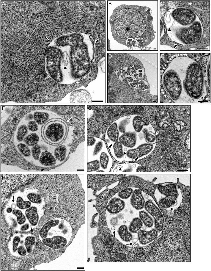
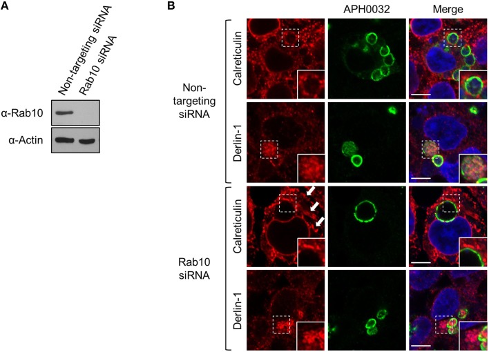
The genus Anaplasma consists of tick-transmitted obligate intracellular bacteria that invade white or red blood cells to cause debilitating and potentially fatal infections. A. phagocytophilum, a human and veterinary pathogen, infects neutrophils to cause granulocytic anaplasmosis. A. marginale invades bovine erythrocytes. Evidence suggests that both species may also infect endothelial cells in vivo. In mammalian and arthropod host cells, A. phagocytophilum and A. marginale reside in host cell derived pathogen-occupied vacuoles (POVs). While it was recently demonstrated that the A. phagocytophilum-occupied vacuole (ApV) intercepts membrane traffic from the trans-Golgi network, it is unclear if it or the A. marginale-occupied vacuole (AmV) interacts with other secretory organelles. Here, we demonstrate that the ApV and AmV extensively interact with the host endoplasmic reticulum (ER) in endothelial, myeloid, and/or tick cells. ER lumen markers, calreticulin, and protein disulfide isomerase, and the ER membrane marker, derlin-1, were pronouncedly recruited to the peripheries of both POVs. ApV association with the ER initiated early and continued throughout the infection cycle. Both the ApV and AmV interacted with the rough ER and smooth ER. However, only derlin-1-positive rough ER derived vesicles were delivered into the ApV lumen where they localized with intravacuolar bacteria. Transmission electron microscopy identified multiple ER-POV membrane contact sites on the cytosolic faces of both species' vacuoles that corresponded to areas on the vacuoles' lumenal faces where intravacuolar Anaplasma organisms closely associated. A. phagocytophilum is known to hijack Rab10, a GTPase that regulates ER dynamics and morphology. Yet, ApV-ER interactions were unhindered in cells in which Rab10 had been knocked down, demonstrating that the GTPase is dispensable for the bacterium to parasitize the ER. These data establish the ApV and AmV as pathogen-host interfaces that directly engage the ER in vertebrate and invertebrate host cells and evidence the conservation of ER parasitism between two Anaplasma species.

Keywords: Anaplasmataceae; Rab; Rickettsia; endoplasmic reticulum; intracellular bacteria; pathogen synapse.


Figures

Figure 1
Figure 1
The ApV engages the host ER throughout A. phagocytophilum infection of mammalian host cells. (A) ER markers localize to and within the ApV in mammalian cells. A. phagocytophilum infected RF/6A cells that had been screened with antibodies against calreticulin or PDI were visualized using LSCM. (B) The ER is recruited to the ApV early and the association is retained throughout the course of infection. RF/6A cells that had been synchronously infected with A. phagocytophilum organisms were screened with antibodies against calreticulin and the pathogen derived ApV membrane protein, APH0032, and examined at several post-infection time points using LSCM. (A,B) Host cell nuclei and bacterial DNA were stained with DAPI (blue). The regions that are demarcated by hatched lined boxes indicate the regions magnified in the insets that are demarcated by solid lined boxes. Scale bars, 5 μm. (C) Percentages of calreticulin-positive ApVs, as identified by DAPI stained intravacuolar A. phagocytophilum bacteria, over the course of a synchronous infection. Data are the means and standard deviations for triplicate samples. (D) Calreticulin cofractionates with A. phagocytophilum organisms. Uninfected (U) or A. phagocytophilum-infected HL-60 cells (I) were homogenized and the post-nuclear supernatants were separated by density gradient centrifugation. Successive one-ml fractions were analyzed by Western blot using antibodies against calreticulin and the A. phagocytophilum major surface protein, P44. Results shown are representative of two experiments with similar results.
Figure 2
Figure 2
Derlin-1-positive ER vesicles are delivered into the ApV lumen where they associate with A. phagocytophilum organisms. (A–D) Derlin-1-positive vesicles are present within the ApV. A. phagocytophilum infected RF/6A cells were screened with antibodies against derlin-1 (A) or derlin-1 and APH0032 (B) and visualized using LSCM. (C) Z-stack series shows derlin-1-labeled vesicles within the ApV. Successive focal planes of the region in (B) that is demarcated by a hatched line box are presented. (D) 3D rendering of the Z-stack series presented in (C) shows derlin-1-positive vesicles encasing A. phagocytophilum organisms within the vacuole. (E) Derlin-1-positive vesicles are recruited to and delivered into the ApV early and continue to detected within the ApV throughout the course of infection. RF/6A cells that had been synchronously infected with A. phagocytophilum organisms were screened with antibodies against derlin-1 and APH0032 and visualized at several post-infection time points using LSCM. (A,E) Regions that are demarcated by hatched lined boxes correspond to the regions magnified in the insets that are demarcated by solid lined boxes. (A–E) Host cell nuclei and bacterial DNA were stained with DAPI (blue). Scale bars, 5 μm. (F) Percentages of ApVs in which derlin-1 signal is detectable within the lumen closely associated with intravacuolar A. phagocytophilum organisms over the course of a synchronous infection. Data are the means and standard deviations for triplicate samples. Results in all panels are representative of two experiments with similar results.
Figure 3
Figure 3
Derlin-1 immunolabeling of intravacuolar A. phagocytophilum bacteria is specific and is reproducible among different derlin-1 antibodies. (A,B) Derlin-1 antibody does not cross-react with any A. phagocytophilum protein. Western blotted lysates of uninfected HL-60 cells and host cell-free A. phagocytophilum (Ap) bacteria (A) or A. phagocytophilum infected derlin-1 siRNA treated (Der1), non-targeting siRNA treated (NT), or uninfected, untreated (U) control HEK-293T cells (B) were probed with antibodies against derlin-1, A. phagocytophilum P44, or ß-actin. (C) Two different derlin-1 antibodies produce comparable immunolabeling patterns of the host cell ER and intravacuolar A. phagocytophilum organisms. A. phagocytophilum and/or uninfected RF/6A cells were screened with derlin-1 antibodies obtained from two different commercial sources. Scale bars, 5 μm. Results shown are representative of two experiments with similar results.
Figure 4
Figure 4
Ectopically overexpressed mCherry-derlin-1 is delivered into and inhibits development of the ApV, and knocking down derlin-1 increases the A. phagocytophilum bacterial load. (A) mCherry-derlin-1 is delivered into the ApV, associated with intravacuolar A. phagocytophilum bacteria, and inhibits ApV development. HEK-293T cells transfected to express mCherry, mCherry-derlin-1, or mock-transfected were incubated with A. phagocytophilum. At 24 h, the cells were fixed, stained with DAPI, and visualized using LSCM. The white arrow in (A) denotes a small mCherry-derlin-1-positive ApV. Scale bars, 0.5 μm. (B) The A. phagocytophilum load is increased in derlin-1 siRNA-treated cells. HEK-293T cells were treated with derlin-1-targeting or non-targeting (NT) siRNA for 72 h. Following siRNA treatment, the cells were infected with A. phagocytophilum for 24 h and total DNA was isolated and subjected to QPCR analysis.
Figure 5
Figure 5
AmVs interact with the ER in mammalian host cells. (A,B) The AmV interacts with the ER and ER-derived vesicles are delivered into its lumen. A. marginale infected RF/6A cells were screened with antibodies against calreticulin (A), PDI (A), or derlin-1 (B). The regions that are demarcated by hatched lined boxes indicate the regions magnified in the insets that are demarcated by solid lined boxes. (C–E) Derlin-1-positive vesicles are present within the AmV in close association with A. marginale bacteria. A. marginale infected RF/6A cells were screened with antibodies against derlin-1 and Msp5 and visualized using LSCM. (D) Z-stack series shows derlin-1-labeled vesicles within the AmV. Successive focal planes of the region in (C) that is demarcated by a hatched line box are presented. (E) 3D rendering of the Z-stack series presented in (D) shows derlin-1-positive vesicles in close proximity to intravacuolar A. marginale organisms. Host cell nuclei and bacteria were stained blue with DAPI. Scale bars, 5 μm. Results shown are representative of two experiments with similar results.
Figure 6
Figure 6
ApVs and AmVs interact with both the RER and SER. Uninfected, A. phagocytophilum infected, or A. marginale infected RF/6A cells were screened with antibodies targeting kinectin-1 or reticulon-4, which are markers for the RER and SER, respectively, and examined using LSCM. Host nuclei and bacterial DNA were stained with DAPI. Scale bars, 5 μm. Results shown are representative of two experiments with similar results.
Figure 7
Figure 7
SIM analyses confirm that calreticulin- and derlin-1-positive vesicles are present in the ApV and AmV lumen in close proximity to intravacuolar Anaplasma spp. bacteria. A. phagocytophilum (A) and A. marginale infected RF/6A cells (B) that had been screened with antibodies against calreticulin or derlin-1 were examined by SIM. The regions in the Merge panels that are demarcated by hatched line boxes indicate the regions that are magnified in the Enlargement panels. Host cell nuclei and bacterial DNA were stained with DAPI (blue). Scale bars, 5 μm. Results shown are representative of two experiments with similar results.
Figure 8
Figure 8
ApVs and AmVs associate with the ER in ISE6 tick cells. Uninfected (C), A. phagocytophilium- (A), or A. marginale-infected ISE6 tick cells (B) were labeled with antibodies against either calreticulin or PDI in combination with APH0032 (for A. phagocytophilum) or Msp2 (for A. marginale) and examined using LSCM. Host nuclei and bacteria were stained blue with DAPI. Scale bars, 5 μm. Results shown are representative of two experiments with similar results.
Figure 9
Figure 9
The ApV interacts with the ER as visualized by TEM. A. phagocytophilum infected HL-60 cells were examined by TEM. White hatched boxes denote areas in the images presented in (B,D) that are presented as enlarged panels in (C,E), respectively. The white arrow in (A) denotes a RER-ApV contact site. Black arrows in (A,C,G) denote autophagosomes contacting the ApV membrane. Black arrowheads in (A,C,G), and (I) demarcate autophagic bodies present within the ApV lumen. White arrowheads in (E) denote ribosomes that label the cytosolic face of the ApV membrane. Thin white arrows in (F,G) demarcate membranes within the ApV lumen associating with A. phagocytophilum organisms. Thin black arrows in (G,H,I) point to vesicles within the ApV lumen in close apposition to A. phagocytophilum bacteria. Scale bars, 0.5 μm. Results shown are representative of two experiments in which a combined total of over 200 different electron micrographs were analyzed.
Figure 10
Figure 10
The AmV interacts with the ER as visualized by TEM. A. marginale infected ISE6 cells were examined by TEM. White hatched boxes denote areas in the images presented in (A,C) that are presented as enlarged panels in (B,D), respectively. The white arrow in (A) denotes a RER-AmV contact site. White arrowheads in (B,D) point to ribosomes that label the cytosolic face of the AmV membrane. Thin black arrows in (A–C) demarcate vesicles within the AmV lumen in close apposition to A. marginale organisms. Scale bars, 0.5 μm. Results shown are representative of two experiments in which a combined total of over 40 different electron micrographs were analyzed.
Figure 11
Figure 11
ApV association with the ER is Rab10-independent. HEK-293T cells were treated with Rab10-targeting or non-targeting siRNA for 72 h. (A) Lysates of non-targeting or Rab10 siRNA treated cells were examined by Western blot for Rab10 knockdown. (B) Following siRNA treatment, the cells were infected with A. phagocytophilum for 48 h, fixed, screened with antibodies against APH0032 and calreticulin or derlin-1, and examined using LSCM. Host cell nuclei and bacterial DNA were stained with DAPI. Regions demarcated by hatched line boxes are magnified in the corresponding inset images that are denoted by a solid line boxes. Arrows point to regions of expansive cisternae, which is characteristic ER morphology in Rab10-depleted cells. Scale bars, 5 μm. Results shown are representative of two experiments with similar results.

Similar articles

Cited by

References

    1. Abu Kwaik Y., Bumann D. (2013). Microbial quest for food in vivo: “nutritional virulence” as an emerging paradigm. Cell. Microbiol. 15, 882–890. 10.1111/cmi.12138 - DOI - PubMed
    1. Agaisse H., Derre I. (2014). Expression of the effector protein IncD in Chlamydia trachomatis mediates recruitment of the lipid transfer protein CERT and the endoplasmic reticulum-resident protein VAPB to the inclusion membrane. Infect. Immun. 82, 2037–2047. 10.1128/IAI.01530-14 - DOI - PMC - PubMed
    1. Allen J. R., Ross S. T., Davidson M. W. (2014). Structured illumination microscopy for superresolution. Chemphyschem 15, 566–576. 10.1002/cphc.201301086 - DOI - PubMed
    1. Al-Zeer M. A., Al-Younes H. M., Kerr M., Abu-Lubad M., Gonzalez E., Brinkmann V., et al. . (2014). Chlamydia trachomatis remodels stable microtubules to coordinate Golgi stack recruitment to the chlamydial inclusion surface. Mol. Microbiol. 94, 1285–1297. 10.1111/mmi.12829 - DOI - PubMed
    1. Beatty W. L. (2006). Trafficking from CD63-positive late endocytic multivesicular bodies is essential for intracellular development of Chlamydia trachomatis. J. Cell Sci. 119, 350–359. 10.1242/jcs.02733 - DOI - PubMed

Publication types

MeSH terms