Review
. 2016 Mar 11:3:6.
doi: 10.1186/s40662-016-0038-6. eCollection 2016.

Assessing progression of keratoconus: novel tomographic determinants

Affiliations
Review

Assessing progression of keratoconus: novel tomographic determinants

Joshua K Duncan et al. Eye Vis (Lond). .

Abstract

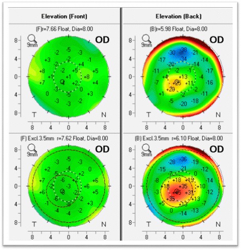
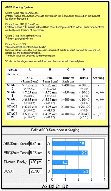
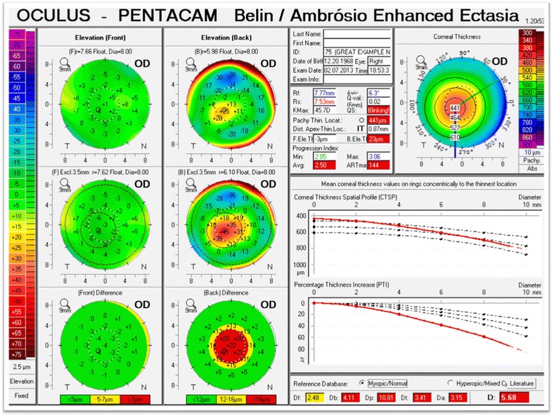
Several methods have been described in the literature to both evaluate and document progression in keratoconus, but there is no consistent or clear definition of ectasia progression. The authors describe how modern corneal tomography, including both anterior and posterior elevation and pachymetric data can be used to screen for ectatic progression, and how software programs such as the Enhanced Reference Surface and the Belin-Ambrosio Enhanced Ectasia Display (BAD) can be employed to detect earlier changes. Additionally, in order to describe specific quantitative values that can be used as progression determinants, the normal noise measurement of the three parameters (corneal thickness at the thinnest point, anterior and posterior radius of curvature (ARC, PRC) taken from the 3.0 mm optical zone centered on the thinnest point), was assessed. These values were obtained by imaging five normal patients using three different technicians on three separate days. The 95 % and 80 % one-sided confidence intervals for all three parameters were surprisingly small (7.88/4.03 μm for corneal thickness, 0.024/0.012 mm for ARC, and 0.083/0.042 mm for PRC), suggesting that they may perform well as progression determinants.

Keywords: Amsler-Krumeich; Collagen cross-linking; Ectatic disease; Keratoconus; Progression; Scheimpflug; Tomography.


Figures

Fig. 1
Fig. 1
Corneal thickness map (left) and Posterior elevation (right). The corneal thickness map shows a thinnest point that is displaced inferiorly and the posterior elevation reveals a prominent posterior island in an eye that has a normal anterior surface (Oculus Pentacam)
Fig. 2
Fig. 2
Scheimpflug optical cross section with edge detection turned on, showing the anterior corneal surface, posterior corneal surface, anterior and posterior lens surfaces identified (Oculus Pentacam)
Fig. 3
Fig. 3
Contralateral eye in a patient with advanced keratoconus in the other eye. The only abnormality seen here (BAD display) is a mild abnormality in the pachymetric progression (Oculus Pentacam)
Fig. 4
Fig. 4
Anterior elevation map (left) showing a prominent paracentral positive island indicative of keratoconus. The map of the left highlights in red the 3.0 mm exclusion zone centered on the thinnest point that is removed from the calculation of the enhanced reference surface
Fig. 5
Fig. 5
Anterior and Posterior elevation maps with the standard BFS (upper maps) and “enhanced reference surface” (lower maps). The standard anterior map (upper left) shows minimal changes against the enhanced reference surface (lower left) as the anterior surface is normal. The standard posterior elevation (upper right) shows an early positive island of elevation that is exaggerated using the enhanced reference surface (lower right) (Oculus Pentacam)
Fig. 6
Fig. 6
The ABCD Keratoconus Grading system currently available on the Topometric/Keratoconus Grading display on the OCULUS Pentacam
Fig. 7
Fig. 7
An example of subclinical keratoconus. The cornea is substantially thinned with a prominent posterior ectasia in spite of a normal anterior surface (BAD display, Oculus Pentacam)

Similar articles

Cited by

References

    1. Nottingham J. Practical Observations on Conical Cornea: and on the Short Sight, and Other Defects of Vision Connected with it. London: J Churchill; 1854.
    1. Rabinowitz YS. Keratoconus. Surv Ophthalmol. 1998;42:297–319. doi: 10.1016/S0039-6257(97)00119-7. - DOI - PubMed
    1. Krachmer JH, Feder RS, Belin MW. Keratoconus and related non- inflammatory corneal thinning disorders. Surv Ophthalmol. 1984;28:293–322. doi: 10.1016/0039-6257(84)90094-8. - DOI - PubMed
    1. Weed KH, McGhee CN, Mac Ewen CJ. Atypical unilateral superior keratoconus in young males. Cont Lens Anterior Eye. 2005;28:177–9. doi: 10.1016/j.clae.2005.10.002. - DOI - PubMed
    1. Gorskova EN, Sevost’ianov EN. Epidemiology of keratoconus in the Urals. Vestn Oftalmol. 1998;114:38–40. - PubMed

LinkOut - more resources