. 2016 Feb;1(2):e85633.
doi: 10.1172/jci.insight.85633. Epub 2016 Feb 25.

Autoimmune response to transthyretin in juvenile idiopathic arthritis

Affiliations

Autoimmune response to transthyretin in juvenile idiopathic arthritis

Cristina C Clement et al. JCI Insight. 2016 Feb.

Abstract

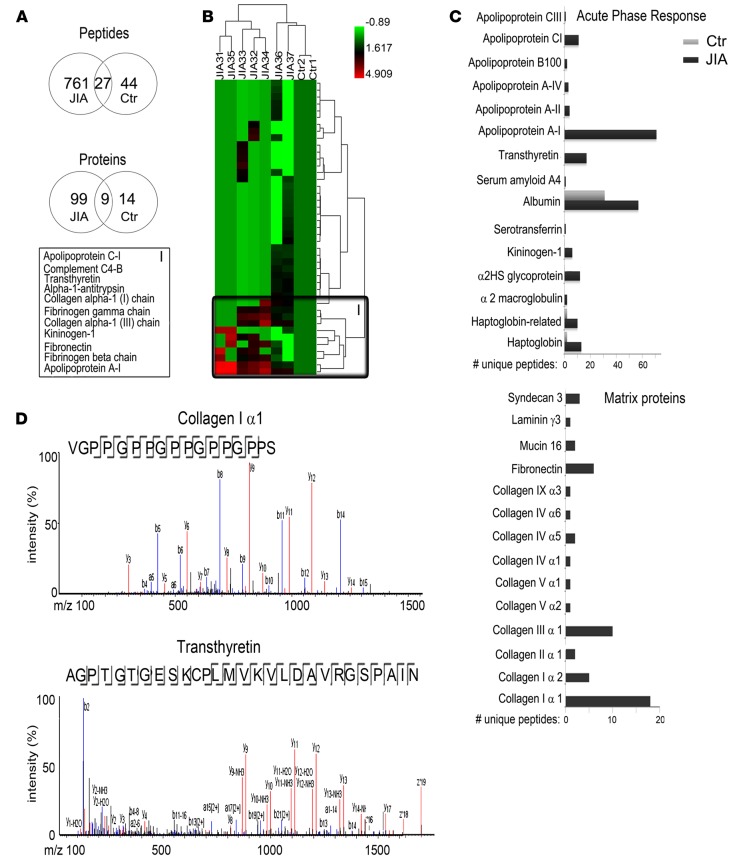
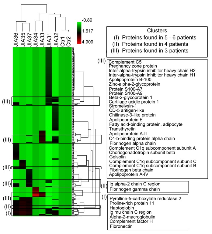
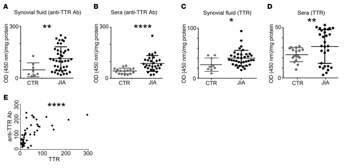
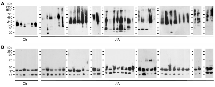
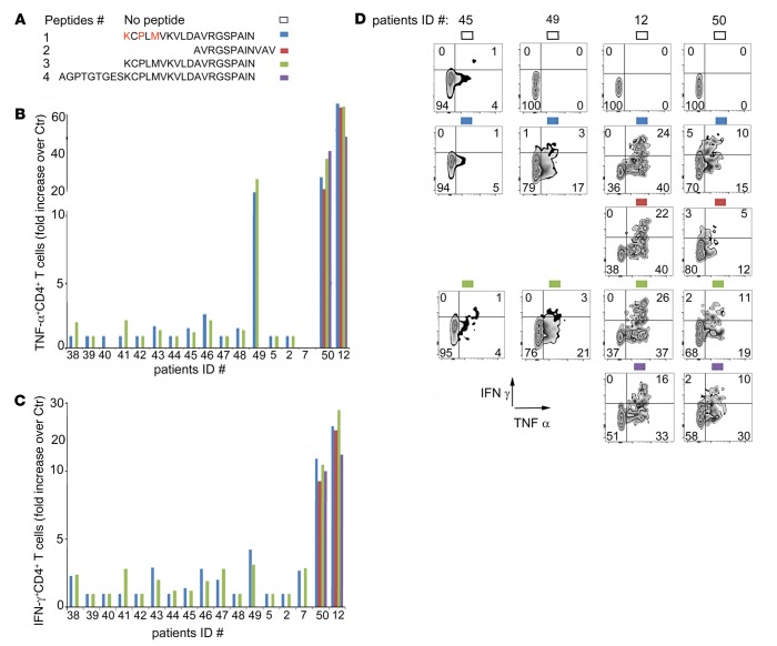
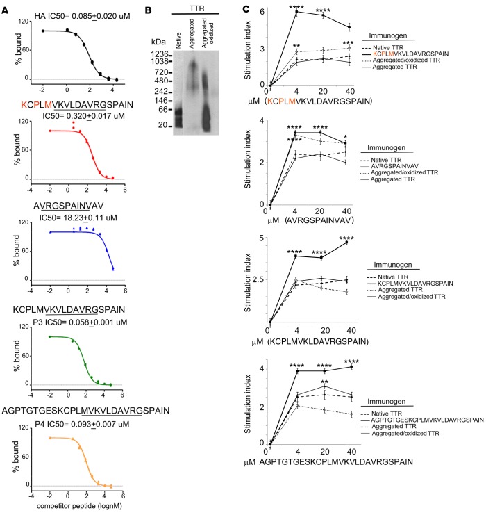
Juvenile idiopathic arthritis (JIA) is the most common pediatric rheumatological condition. Although it has been proposed that JIA has an autoimmune component, the autoantigens are still unknown. Using biochemical and proteomic approaches, we identified the molecular chaperone transthyretin (TTR) as an antigenic target for B and T cell immune responses. TTR was eluted from IgG complexes and affinity purified from 3 JIA patients, and a statistically significant increase in TTR autoantibodies was observed in a group of 43 JIA patients. Three cryptic, HLA-DR1-restricted TTR peptides, which induced CD4+ T cell expansion and IFN-γ and TNF-α production in 3 out of 17 analyzed patients, were also identified. Misfolding, aggregation and oxidation of TTR, as observed in the synovial fluid of all JIA patients, enhanced its immunogenicity in HLA-DR1 transgenic mice. Our data point to TTR as an autoantigen potentially involved in the pathogenesis of JIA and to oxidation and aggregation as a mechanism facilitating TTR autoimmunity.


Figures

Figure 1
Figure 1. Peptidomic profiling of synovial fluid from juvenile idiopathic arthritis patients and controls.
(A) Venn diagram reporting the number of endogenous peptides and their protein substrates (peptidomic data are reported in Supplemental Tables 2 and 3) found in the synovial fluid of patients with juvenile idiopathic arthritis (JIA) and controls (data were compiled from 3 separate proteomic analyses). (B) Hierarchical clustering analysis of the protein substrates of the peptidome found in the synovial fluid of patients with JIA and controls was generated in Scaffold 4 using the log of the normalized spectral abundance factor [ln(NSAF)] values [average of 3 experiments for the ln(NSAF)]. The Ward’s dual-clustering method and the t test were performed comparing 2 controls and 7 JIA patients. Only 43 protein substrates of the peptidome were shown to have more than 1.5-fold difference in expression across all patients, with statistical significance corresponding to a FDR < 0.7 [green corresponds to the lowest ln(NSAF), while red corresponds to the highest ln(NSAF)]. The protein substrates with the highest statistically significant contribution to the peptidome of JIA patients versus controls are shown as cluster (I) (at P < 0.08) (peptidomic data used to compile the heat map are reported in Supplemental Table 2F). (C) Analysis of the unique endogenous peptides derived from the degradation of the acute-phase response and cartilage matrix proteins found in the synovial fluid of patients with JIA and controls (data were compiled from 3 separate proteomic analyses). The enhanced degradation of the proteome from the synovial fluid of JIA patients correlates with the increased number of sequenced endogenous peptides. (D) Selected tandem mass spectrometry fragmentation profiles of peptides derived from collagen I (α 1) and from transthyretin, as mapped in the synovial fluid of patients with JIA.
Figure 2
Figure 2. Degradome and posttranslational modifications associated with the peptidome found in the synovial fluid of patients with juvenile idiopathic arthritis and controls.
(A) Number of peptide cleavage sites by each specified protease as analyzed by MEROPS and CutDB, found in the synovial fluid of patients with juvenile idiopathic arthritis (JIA) and controls (data were compiled from 3 separate proteomic analyses). (B) Posttranslational modifications (PTMs), mapped on the sequenced peptides, present in the synovial fluid of patients with JIA (n = 7) and controls (n = 2). Pie charts illustrate the percentage of posttranslational modified peptides found in the synovial fluid of patients with JIA and controls (data were compiled from 3 separate proteomic analyses). (C) Quantification of the increased carbonyl content in the proteome of the synovial fluid from JIA patients as compared with controls. Equal amounts of total protein from the synovial fluid of each JIA patient and control (10–20 μg) were derivatized with the reagent dinitrophenylhydrazine (DNPH), and the total carbonyl content (as nanomoles/mg protein) was determined spectroscopically. Average readings (mean ± SD) from 3 separate experiments are shown as bar graphs. Data were analyzed by 1-way ANOVA (P < 0.005) followed by Tukey test.
Figure 3
Figure 3. IgG-eluted immune proteome from the synovial fluid of patients with juvenile idiopathic arthritis and controls.
Hierarchical cluster analysis (HCA) of proteins eluted from the IgG purified from the synovial fluid of patients with juvenile idiopathic arthritis (JIA) and controls (data were compiled from 3 separate proteomic analyses). Quantitative data sets were generated for each protein entry in Scaffold 4 using the log of the normalized spectral abundance factor [ln(NSAF)] values [average of 3 experiments for the ln(NSAF)]. The Ward’s dual-clustering method followed by the t test was performed for comparison of 2 controls and 7 JIA patients. The HCA depicts proteins with at least a 1.5-fold difference in expression between the JIA and the control groups, with statistical significance corresponding to a FDR value < 0.7 [green corresponds to the lowest ln(NSAF), while red corresponds to the highest ln(NSAF)] (proteomic data used to compile the heat map are reported in Supplemental Table 5).
Figure 4
Figure 4. Transthyretin and anti-transthyretin antibodies in the synovial fluid and sera of juvenile idiopathic arthritis patients and controls.
(A) Quantification of transthyretin (TTR) autoantibodies, as detected by ELISA, present in the synovial fluid from patients with juvenile idiopathic arthritis (JIA) and controls (data were compiled from 3 separate ELISAs, with each patient’s samples run in quadruplicate). Data were analyzed by 1-way ANOVA and Tukey test. (B) Quantification of TTR autoantibodies, as detected by ELISA, present in the sera of patients with JIA and controls (data were compiled from 3 separate ELISAs, with each patient’s samples run in quadruplicate). Data were analyzed by 1-way ANOVA and Tukey test. (C and D) Amount of TTR present in the synovial fluid and sera from patients with JIA and controls (data were compiled from 3 separate ELISAs, with each patient’s samples run in quadruplicate) Data were analyzed by 1-way ANOVA and Tukey test. (E) Correlation between TTR protein and anti-TTR antibodies (Spearman r = 0.72, ****P < 0.001). The patient ID for each circle is reported in Supplemental Figure 4. Mean ± SD. *P < 0.05; **P < 0.005; ****P < 0.001.
Figure 5
Figure 5. Transthyretin aggregates in the synovial fluid of juvenile idiopathic arthritis.
Western blot analysis of transthyretin (TTR) proteins present in the synovial fluid of controls and juvenile idiopathic arthritis (JIA) patients run on a (A) native gel or (B) SDS-PAGE (representative gel out of 2 runs). TTR monomers (15 kDa), dimers (30 kDa), tetramers (60 kDa), and aggregates (above 60 kDa) are visible in JIA patients when run on a native gel. The patient ID for each lane is reported in Supplemental Figure 5.
Figure 6
Figure 6. Production of TNF-α and IFN-γ by T cells isolated from synovial fluid of juvenile idiopathic arthritis patients in response to transthyretin peptides.
(A) Primary sequence of transthyretin (TTR) peptides. Residues in red are posttranslational modified (M is oxidized, and K and P are hydroxylated). (B) Fold increase of CD4+ T cells producing TNF-α found in the synovial fluid (SF) of juvenile idiopathic arthritis (JIA) patients. Data are reported as fold increase in the number of TNF-α–positive cells following stimulation with TTR peptides over unstimulated cells (1 out of 2 representative experiments is shown). (C) Fold increase of CD4+ T cells producing IFN-γ found in the SF of JIA patients (one out of two representative experiments is shown). Data are reported as fold increase in the number of IFN-γ–positive cells following stimulation with TTR peptides over unstimulated cells. (D) Examples of FACS analysis of TNF-α– and IFN-γ–producing T cells from different JIA patients before and after stimulation with TTR peptides. HLA-DR type of patient 12 is DRB1*03:07/07:01 and that of patient 50 is DRB1*01:01/11:01.
Figure 7
Figure 7. Proliferative responses of T cells isolated from synovial fluid of juvenile idiopathic arthritis patients after stimulation with transthyretin peptides.
Histograms of CFSE-labeled T cells from different juvenile idiopathic arthritis (JIA) patients before and after stimulation with transthyretin peptides (1 out of 2 representative experiments is shown). HLA-DR type of patient 12 is DRB1*03:07/07:01 and that of patient 50 is DRB1*01:01/11:01.
Figure 8
Figure 8. HLA-DR1–binding affinity and proliferative response to transthyretin peptides following immunization of HLA-DR1 mice with native, aggregated, or aggregated/oxidized transthyretin.
(A) Inhibition binding curves (percentage bound test peptide vs. test peptide concentration [nM]) for transthyretin (TTR) peptides. Binding to HLA-DR1 molecules measured at 72 hours (1 out of 3 representative experiments is shown). Shown at the top of the graphs are the peptide sequences as well as the calculated values for binding (IC50 [μM]). Binding of the immunodominant viral epitope HA peptide is shown as control. (B) Native gel showing native TTR (monomer at 15 kDa and tetramer at 60 kDa); aggregated TTR, following exposure to low pH; and aggregated and oxidized TTR, following oxidation by Fenton reaction. One out of three gels is shown. (C) T cell–proliferative responses to increasing concentrations of the reported TTR peptides following immunization with native or aggregated or aggregated/oxidized TTR. HLA-DR1 mice were immunized as reported in Methods, popliteal and axillary nodes were harvested 3 weeks later, and T cells were rechallenged in vitro with increasing concentrations of the reported immunogens. Data are reported as stimulation index (BrDu incorporation following antigen stimulation over BrDu incorporation in absence of specific antigen). Data were compiled from 3 separate immunizations (n = 6). Experiments to evaluate T cell proliferation in response to antigen were run in quadruplicate for each antigen concentration tested. Mean ± SD. Data were analyzed by 1-way ANOVA (P < 0.005) and Tukey test. Asterisks indicate statistical significance, calculated at each concentration, between the peptide stimulation index and the native TTR or between the native TTR and the unfolded/carbonylated TTR. *P < 0.05; **P < 0.01; ***P < 0.005; ****P < 0.001.

Similar articles

Cited by

References

    1. Alexeeva EI, et al. Efficacy and safety of repeat courses of rituximab treatment in patients with severe refractory juvenile idiopathic arthritis. Clin Rheumatol. 2011;30(9):1163–1172. doi: 10.1007/s10067-011-1720-7. - DOI - PubMed
    1. Barnes MG, et al. Biologic similarities based on age at onset in oligoarticular and polyarticular subtypes of juvenile idiopathic arthritis. Arthritis Rheum. 2010;62(11):3249–3258. doi: 10.1002/art.27657. - DOI - PMC - PubMed
    1. Finnegan S, et al. Synovial membrane protein expression differs between juvenile idiopathic arthritis subtypes in early disease. Arthritis Res Ther. 2014;16(1):e85633. doi: 10.1186/ar4434. - DOI - PMC - PubMed
    1. Gibson DS, et al. Proteomic analysis of recurrent joint inflammation in juvenile idiopathic arthritis. J Proteome Res. 2006;5(8):1988–1995. doi: 10.1021/pr060129t. - DOI - PubMed
    1. Gibson DS, Qiu J, Mendoza EA, Barker K, Rooney ME, LaBaer J. Circulating and synovial antibody profiling of juvenile arthritis patients by nucleic acid programmable protein arrays. Arthritis Res Ther. 2012;14(2):e85633. doi: 10.1186/ar3800. - DOI - PMC - PubMed