. 2016 Aug;55(2):288-98.
doi: 10.1165/rcmb.2015-0197OC.

Oxidized Ferric and Ferryl Forms of Hemoglobin Trigger Mitochondrial Dysfunction and Injury in Alveolar Type I Cells

Affiliations

Oxidized Ferric and Ferryl Forms of Hemoglobin Trigger Mitochondrial Dysfunction and Injury in Alveolar Type I Cells

Narendranath Reddy Chintagari et al. Am J Respir Cell Mol Biol. 2016 Aug.

Abstract

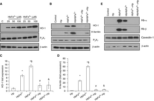
Lung alveoli are lined by alveolar type (AT) 1 cells and cuboidal AT2 cells. The AT1 cells are likely to be exposed to cell-free hemoglobin (Hb) in multiple lung diseases; however, the role of Hb redox (reduction-oxidation) reactions and their precise contributions to AT1 cell injury are not well understood. Using mouse lung epithelial cells (E10) as an AT1 cell model, we demonstrate here that higher Hb oxidation states, ferric Hb (HbFe(3+)) and ferryl Hb (HbFe(4+)) and subsequent heme loss play a central role in the genesis of injury. Exposures to HbFe(2+) and HbFe(3+) for 24 hours induced expression of heme oxygenase (HO)-1 protein in E10 cells and HO-1 translocation in the purified mitochondrial fractions. Both of these effects were intensified with increasing oxidation states of Hb. Next, we examined the effects of Hb oxidation and free heme on mitochondrial bioenergetic function by measuring changes in the mitochondrial transmembrane potential and oxygen consumption rate. In contrast to HbFe(2+), HbFe(3+) reduced basal oxygen consumption rate, indicating compromised mitochondrial activity. However, HbFe(4+) exposure not only induced early expression of HO-1 but also caused mitochondrial dysfunction within 12 hours when compared with HbFe(2+) and HbFe(3+). Exposure to HbFe(4+) for 24 hours also caused mitochondrial depolarization in E10 cells. The deleterious effects of HbFe(3+) and HbFe(4+) were reversed by the addition of scavenger proteins, haptoglobin and hemopexin. Collectively, these data establish, for the first time, a central role for cell-free Hb in lung epithelial injury, and that these effects are mediated through the redox transition of Hb to higher oxidation states.

Keywords: heme oxygenase; hemoglobin; hemolysis; lung epithelium; mitochondria.


Figures

Figure 1.
Figure 1.
Representative spectra of ferrous hemoglobin (HbFe2+), ferric Hb (HbFe3+), and ferryl Hb (HbFe4+): Hb was isolated from human blood. HbFe3+ was prepared as described in Materials and Methods. HbFe4+ was prepared by adding 10-molar excess of hydrogen peroxide (H2O2) to HbFe3+. The HbFe4+ was derivatized by addition of Na2S to produce sulfHb (absorption peak at 620 nm). A spectral scan was performed from 450 to 700 nm to confirm oxidation status and quantify the heme content. Shown are representative spectra (n > 6). The spectra of different Hb oxidation states are identified by characteristic peaks in the visible region (HbFe2+, 541 and 576 nm; HbFe3+, 500 and 630 nm; HbFe4+, 544 nm, 585 nm, and a flattened region between 600 and 700 nm). AU, arbitrary units.
Figure 2.
Figure 2.
Haptoglobin (Hp) attenuates HbFe2+- and HbFe3+-induced effects in mouse lung epithelial (E10) cells: E10 cells were exposed to HbFe2+ and HbFe3+ at the indicated concentration for 24 hours. The cell lysates were immunoblotted to confirm the expression of heme oxygenase (HO)-1 and P2X7 receptor proteins. Equal loading was confirmed by reprobing the blots against β-actin. A dose-dependent increase was observed after exposure to HbFe2+ and HbFe3+. (A) A representative Western blot image after exposure to increasing doses of HbFe2+ and HbFe3+. The E10 cells were exposed to human HbFe2+ (50 μM) and HbFe3+ (50 μM) in the presence and absence of Hp (50 μM) for 24 hours. The cell lysates were probed for expression of HO-1 and H-ferritin proteins. Equal loading was confirmed by reprobing the blots with β-actin. (B) Representative images of HO-1, H-ferritin, and P2X7 receptor (n = 5). The expression of HO-1 and H-ferritin was normalized against β-actin. Fold expressions are shown for (C) HO-1 and (D) H-ferritin. The cell lysates were probed for detection of human Hb α and β subunits in the cell lysates. The Western blot images are shown in E (n = 4). The treatment groups were compared using unpaired Student’s t test. *P < 0.05 versus control; $P < 0.05 versus HbFe2+; #P < 0.05 versus HbFe2+; and &P < 0.05 versus HbFe3+. Data presented are means (±SEM) of fold expression.
Figure 3.
Figure 3.
HbFe3+ up-regulates mitochondrial HO-1 expression and alters basal mitochondrial respiration: mouse E10 cells were exposed to HbFe2+ and HbFe3+ with and without Hp for 24 hours. The mitochondria were isolated and probed for expression of HO-1 by Western blotting. The blots were reprobed with cytochrome c oxidase IV (COX-IV), a mitochondrial marker protein. The expression of glyceraldehyde phosphate dehydrogenase (GAPDH) and extracellular signal–regulated kinase (ERK) (as possible cytosolic markers) was also used to confirm the purity of the mitochondria. (A) Shown are representative images (n = 3). Mitochondrial HO-1 levels were quantified as fold expression over control after normalization with COX IV (n = 3). (B) Shown are means (±SEM) of fold expression. Mitochondrial function was analyzed by extracellular flux analysis. Various bioenergetics parameters were calculated from the shown oxygen consumption rate (OCR) plots. Shown is (C) OCR plot and (D) bioenergetic profile of E10 cells after exposure to HbFe2+ and HbFe3+ in the presence and absence of Hp. OCR plot shown is from one representative experiment repeated three times. *P < 0.05 versus control; #P < 0.05 versus HbFe3+, t test, unpaired. AA, antimycin; FCCP, carbonyl cyanide-4-(trifluoromethoxy)phenylhydrazone; Oligo, oligomycin; Rot, rotenone.
Figure 4.
Figure 4.
HO-1 translocates into mitochondria after exposure to oxidized Hbs: E10 cells were exposed to freshly prepared HbFe4+ for 12 hours in the presence and absence of equimolar concentration of Hp. (A) Representative immunoblot images are shown. The expression of HO-1 was quantified by comparison with β-actin. (B) Shown is fold expression of HO-1. Mitochondria (and cytosolic fractions) were isolated and probed for the expression of HO-1 and cytosolic markers. *P < 0.05 versus Hp; $P < 0.05 versus HbFe2+; #P < 0.05 versus HbFe4+. (C) Shown are the representative immunoblot images. The expression and localization of HO-1 was also confirmed by confocal microscopy. (D) Shown are representative confocal images indicating expression of HO-1 and Mitotracker Red. Data presented are means (±SEM). Con, control.
Figure 5.
Figure 5.
Oxidized Hbs alter mitochondrial membrane potential and cause dysfunction: E10 cells were exposed to HbFe2+, HbFe3+, and freshly prepared HbFe4+ for a period of 12 hours (for XF24 assay) or 24 hours (for membrane potential assay) in the presence and absence of equimolar concentration of Hp. The mitochondrial membrane potential was measured after incubation using tetraethyl-benzimidazolyl carbocyanine iodide (JC-1) dye. (A) Shown is the JC-1 590:530-nm emission ratio expressed as a percent of oligomycin-treated hyperpolarized cells. The cells were exposed to hemin (5 and 10 µM), HbFe2+, and HbFe3+ as described earlier. The cell lysates were probed for expression of HO-1. Equal loading was confirmed by reprobing the immunoblots against β-actin. Shown in the upper panel is a representative immunoblot. The mitochondrial oxygen consumption was measured using XF24 extracellular flux analyzer. (B) OCR plot after exposure to HbFe4+ (n = 4). (C) Bioenergetic profile was calculated from the representative OCR plot repeated three times. *P < 0.05 versus control; #P < 0.05 versus HbFe4+, t test, unpaired. Data presented are means (±SEM).
Figure 6.
Figure 6.
Proposed mechanism of Hb (and its oxidized species)-induced toxicity: Hb undergoes oxidation to higher oxidation states, such as HbFe3+ and HbFe4+. These products activate the NF-κB pathway, leading to release of chemokines. The oxidized species lose heme-iron easily, leading to increased HO-1 and ferritin synthesis. HO-1 translocates into the mitochondria, leading to possible dysfunction. Free heme and CO generated by HO-1 can also inhibit mitochondrial respiratory chain function (electron transport chain [ETC]/mitochondrial transmembrane potential [ΔΨm]). Hp attenuates the Hb-induced effects, possibly by diffusing Hb radicals. Caveoli and some unknown pathways may also be involved in endocytosis of Hb. BV, biliverdin; HBOC, Hb-based oxygen carrier; Hpx, hemopexin; TLR4, Toll-like receptor 4.

References

    1. Reeder BJ. The redox activity of hemoglobins: from physiologic functions to pathologic mechanisms. Antioxid Redox Signal. 2010;13:1087–1123. - PubMed
    1. Jia Y, Wood F, Buehler PW, Alayash AI. Haptoglobin preferentially binds β but not α subunits cross-linked hemoglobin tetramers with minimal effects on ligand and redox reactions. PLoS One. 2013;8:e59841. - PMC - PubMed
    1. Schaer DJ, Schaer CA, Buehler PW, Boykins RA, Schoedon G, Alayash AI, Schaffner A. CD163 is the macrophage scavenger receptor for native and chemically modified hemoglobins in the absence of haptoglobin. Blood. 2006;107:373–380. - PubMed
    1. Kristiansen M, Graversen JH, Jacobsen C, Sonne O, Hoffman HJ, Law SK, Moestrup SK. Identification of the haemoglobin scavenger receptor. Nature. 2001;409:198–201. - PubMed
    1. Gozzelino R, Jeney V, Soares MP. Mechanisms of cell protection by heme oxygenase-1. Annu Rev Pharmacol Toxicol. 2010;50:323–354. - PubMed

Publication types

MeSH terms