. 2016 Mar 14;11(3):e0150555.
doi: 10.1371/journal.pone.0150555. eCollection 2016.

Axial Spondylometaphyseal Dysplasia Is Caused by C21orf2 Mutations

Affiliations

Axial Spondylometaphyseal Dysplasia Is Caused by C21orf2 Mutations

Zheng Wang et al. PLoS One. .

Abstract

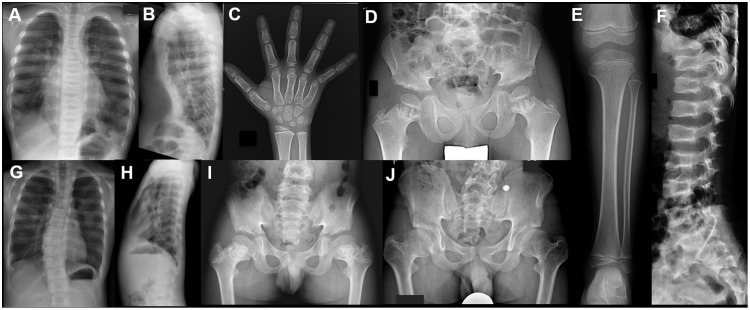
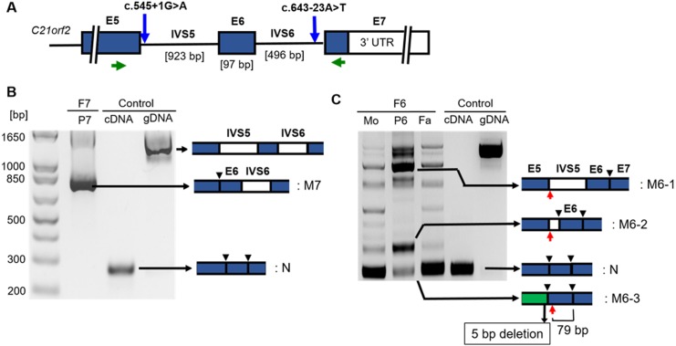
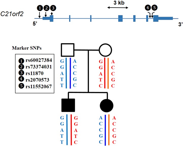
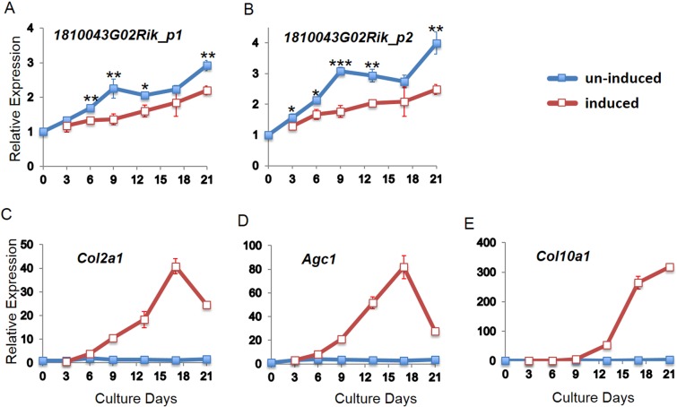
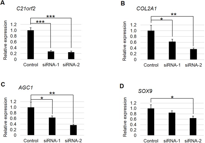
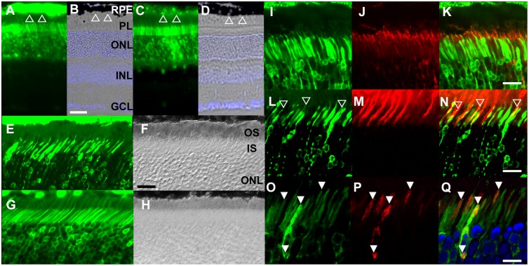
Axial spondylometaphyseal dysplasia (axial SMD) is an autosomal recessive disease characterized by dysplasia of axial skeleton and retinal dystrophy. We conducted whole exome sequencing and identified C21orf2 (chromosome 21 open reading frame 2) as a disease gene for axial SMD. C21orf2 mutations have been recently found to cause isolated retinal degeneration and Jeune syndrome. We found a total of five biallelic C21orf2 mutations in six families out of nine: three missense and two splicing mutations in patients with various ethnic backgrounds. The pathogenic effects of the splicing (splice-site and branch-point) mutations were confirmed on RNA level, which showed complex patterns of abnormal splicing. C21orf2 mutations presented with a wide range of skeletal phenotypes, including cupped and flared anterior ends of ribs, lacy ilia and metaphyseal dysplasia of proximal femora. Analysis of patients without C21orf2 mutation indicated genetic heterogeneity of axial SMD. Functional data in chondrocyte suggest C21orf2 is implicated in cartilage differentiation. C21orf2 protein was localized to the connecting cilium of the cone and rod photoreceptors, confirming its significance in retinal function. Our study indicates that axial SMD is a member of a unique group of ciliopathy affecting skeleton and retina.


Conflict of interest statement

Competing Interests: The authors have declared that no competing interests exist.

Figures

Fig 1
Fig 1. Radiographic features of axial SMD.
(A-F) P7 at age 6 years. Note narrow thorax, short ribs with cupped anterior ends, mildly serrated iliac wings, short ilia, metaphyseal irregularities and shortening of the proximal femora, and mild platyspondyly. Metaphyses of knee and ankle are normal. Hands are normal. G-I) P5 at age 10 years. Narrow thorax with short ribs, mildly serrated iliac wings, short ilia, and metaphyseal irregularities and shortening of the proximal femora. He had mild scoliosis, but platyspondyly is not evident. J) P5 at age 14 years. Note progressive shortening and varus deformity of the proximal femora.
Fig 2
Fig 2. Analysis of the splicing mutations.
(A) A schematic of the local genomic structure of C21orf2. Positions of the splicing donor site mutation (c.545+1G>A) in F6 and the branch-point mutation (c.643-23A>T) in F1 and F7 are indicated by blue arrows. E: exon, IVS: intron, Green arrows: positions of RT-PCR primers. B) RT-PCR analysis for c.643-23A>T. Intron 6 was not spliced in the mutant transcript (M7), which had a frame shift with the elongated reading frame. N: normal transcript. Black arrowhead: splicing junction in specific transcript. C) RT-PCR analysis for c.545+1G>A. In the family members (F6), aberrant bands with various sizes (M6-1~3) were obtained. Sanger-sequencing revealed that M6-3, an apparently normal size band in the patient (P6) represented a miss-spliced mutant which lost 5 bp in the end of exon 5. Red arrow: position of the stop codon. In M6-1 and 3, the new stop codons are more than 55 bp upstream of the last splicing junctions. In M6-2, the new stop codon is in the 3rd last exon. Therefore, all these transcripts are considered to receive nonsense-mediated mRNA decay. Mo: the mother; Fa: the father.
Fig 3
Fig 3. A haplotype analysis of C21orf2 in family F3.
The sib patients inherited different C21orf2 haplotypes from the parents, respectively, which ruled out C21orf2 as a disease gene in this family.
Fig 4
Fig 4. C21orf2 expression during chondrocyte induction.
Relative mRNA expression of mouse C21orf2 (1810043G02Rik) in induced (red lines) and un-induced (blue lines) ATDC5 cells. (A-B) The expression of 1810043G02Rik measured by real-time PCR using two primer sets; (C-E) Expression of chondrocyte marker genes (Col2a1, Agc1 and Col10a1), indicating the differentiation of induced ATDC5 cell to chondrocyte. All the expression values were presented relatively to the ones of day 0, which was set as 1. *: P< 0.05, **: P< 0.01, ***: P< 0.001; induced versus un-induced by t-test. n = 3.
Fig 5
Fig 5. Effects of siRNAs for C21orf2 on chondrocyte marker genes in OUMS-27 cell.
(A) C21orf2 was significantly knocked-down by both siRNAs (siRNA-1 and 2). (B-D) mRNA expression of chondrocyte differentiation marker genes. The expression of the marker genes decreased when C21orf2 was knocked-down. *: P< 0.05, **: P< 0.01, ***: P< 0.001; versus control by t-test. n = 3.
Fig 6
Fig 6. C21orf2 localized to the connecting cilium of the rod and cone photoreceptors.
(A-D) Expression of EGFP driven by CMV-promoter or C21orf2-promoter. When driven by the ubiquitous CMV-promoter, EGFP showed stronger expression in the retinal pigment epithelium (RPE; Open triangle) than in the photoreceptors (A, B). When driven by the C21orf2-promoter, EGFP is expressed more prominently in photoreceptors than in RPE (C, D). (E-H) AAV8-mediated expression of EGFP fusion protein. The C21orf2-EGFP fusion protein was not detected in the outer segments (OS; E, F), while EGFP was present in the OS in the control (G, H). (I-K) C21orf2 localized to the connecting cilium (red; stained with anti-acetylated-tubulin antibodies). (L-N) Association of C21orf2 to the connecting cilium, but not to the surrounding OS structure in cone photoreceptors. C21orf2-EGFP fusion protein remains localized to the cilia (open arrowheads) inside the PNA-positive cone OS (red). (O-Q) Lack of spatial association between C21orf2 and mitochondria. Kusabira Orange-tagged mitochondria (red). RPE, retinal pigment epithelium; PL, photoreceptor layer; ONL, outer nuclear layer; INL, inner nuclear layer; GCL, ganglion cell layer; OS, outer segment; IS inner segment. Scales bars: 50 μm (B), 30 μm (H, K, N) and 15 μm (Q).

References

    1. Warman ML, Cormier-Daire V, Hall C, Krakow D, Lachman R, Lemerrer M, et al. Nosology and classification of genetic skeletal disorders: 2010 revision. American Journal of Medical Genetics, Part A. 2011. pp. 943–968. 10.1002/ajmg.a.33909 - DOI - PMC - PubMed
    1. Ehara S, Kim OH, Maisawa S, Takasago Y, Nishimura G. Axial spondylometaphyseal dysplasia. Eur J Pediatr. 1997;156: 627–630. 10.1007/s004310050679 - DOI - PubMed
    1. Isidor B, Baron S, Van Kien PK, Bertrand AM, David A, Le Merrer M. Axial spondylometaphyseal dysplasia: Confirmation and further delineation of a new SMD with retinal dystrophy. Am J Med Genet Part A. 2010;152: 1550–1554. 10.1002/ajmg.a.33397 - DOI - PubMed
    1. Suzuki S, Kim OH, Makita Y, Saito T, Lim GY, Cho TJ, et al. Axial spondylometaphyseal dysplasia: Additional reports. Am J Med Genet Part A. 2011;155: 2521–2528. 10.1002/ajmg.a.34192 - DOI - PubMed
    1. Khan AO, Eisenberger T, Nagel-wolfrum K, Wolfrum U, Bolz HJ. C21orf2 is mutated in recessive early-onset retinal dystrophy with macular staphyloma and encodes a protein that localises to the photoreceptor primary cilium. 2015; 1725–1731. 10.1136/bjophthalmol-2015-307277 - DOI - PubMed

MeSH terms

Supplementary concepts