. 2016 Mar;57(3):1153-9.
doi: 10.1167/iovs.15-17753.

Anatomical Variation of Human Collector Channel Orifices

Affiliations

Anatomical Variation of Human Collector Channel Orifices

Michael D Bentley et al. Invest Ophthalmol Vis Sci. 2016 Mar.

Abstract

Purpose: To examine the anatomical variation of normal human collector channel orifices and their relationship with Schlemm's canal.

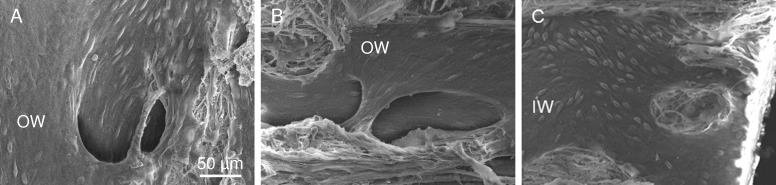
Methods: Ten human anterior segments fixed by immersion or perfusion were dissected radially and further divided by fine dissection into corresponding inner and outer wall segments. The tissues were dehydrated, critical-point dried, sputter coated, and examined by scanning electron microscopy. Images were obtained at magnifications from ×200 to ×10,000. Selected radial collector channel regions were processed for plastic embedding.

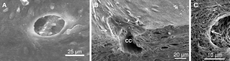
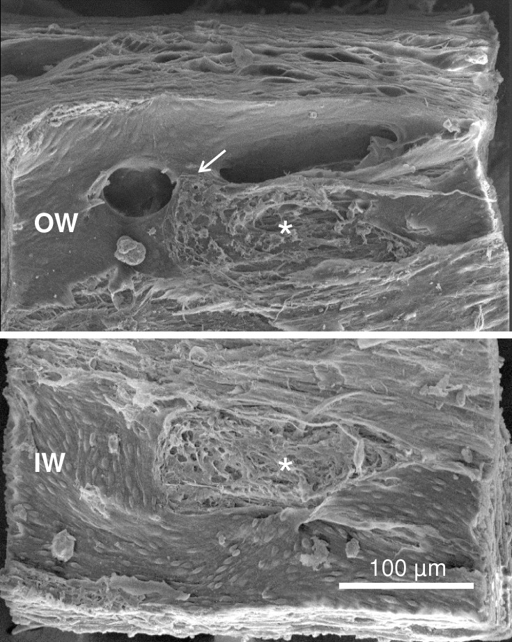
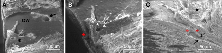
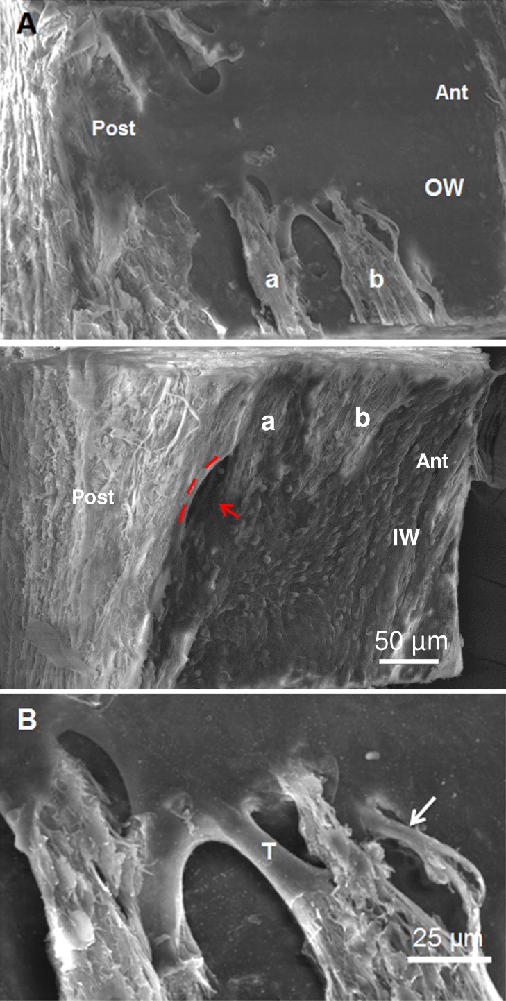
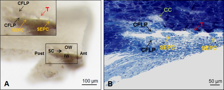
Results: Two classes of collector channel orifices were identified. Simple oval orifices (54.7 ± 4.6-μm diameter) were lined with endothelial cells and most often occurred on a planar region of Schlemm's canal outer wall. Complex orifices (62.7 ± 3.4-μm diameter) were often found associated with septal columns and bridges, and typically covered with flap-like structures (10-40 μm) that extended between the inner and outer wall and over the collector channel orifices. Both simple and complex orifices had complete or partial lip-like rims. In orifices with partial rims, a trough-like groove was often visible on the outer wall surface opposite the lip. Transected septa and inner and outer wall adhesion sites were often found in association with complex collector channel orifices.

Conclusions: Collector channel orifice structure varied from simple ovals to complex tethered flaps and bridges. Collector channel orifices with complex flaps connect the inner and outer walls of Schlemm's canal, and may serve to enhance and regulate aqueous outflow in these regions.


Figures

Figure 1
Figure 1
Various types of simple collector channel (CC) orifices in the outer wall of Schlemm's canal. (A) Representative image showing a simple orifice with thickened rim surrounding entire CC opening. (B) Transected orifice showing lip-like rim (white arrow) in association with a CC. (C) Underlying matrix of orifice rim denuded of endothelium. Arrow points to circumferential fibers at orifice rim.
Figure 2
Figure 2
Complex orifice with bridge. Elongated orifice with overlying bridge (arrow). Outer wall (OW, top) and inner wall (IW, bottom) are joined at corresponding oval adhesion sites (asterisks). Inner wall cells appeared rounded and elevated in contrast to OW cells that appeared elongated and flat.
Figure 3
Figure 3
Complex CC showing relationship to adjacent simple CC, flap covering orifice and intrascleral CC. (A) Low power of OW showing complex orifice with flap (asterisk) and simple orifice (arrow). (B) Higher magnification of complex flap with endothelial cells covering its surface (asterisk) and two flap “anchors” extending from its edge (arrowheads). Note on the longer flap a collagenous core is visible. Flap covers entrance to CC in the sclera (red arrow head) and distally extends from the orifice 175 μm. (C) Side view showing the orifice neck (red arrowheads) and CC proper (black arrows). Complex flap is visible over the CC orifice in the OW with extension into the CC proper in the sclera.
Figure 4
Figure 4
Complex CC orifice associated with flaps. (A) Corresponding OW (top) and IW (bottom) showing flap-structure on OW overlying orifice at the junctional region of the IWs and OWs (red arrow). Lip of the orifice (IW panel, middle) is delineated by three red dashes on the top and a red arrow, which points to the darkened orifice region. Extensions and corresponding anchor sites on IWs and OWs are labeled a and b. Anterior and posterior are labeled in OW and IW wedges. (B) Enlargement of Figure 4A OW (top) near region labelled “b” showing underlying fibrous structure of anchoring tether (T) and tubule-like structure (arrow).
Figure 5
Figure 5
Gross dissection of complex flap in Schlemm's canal and correlative plastic section. (A) Gross dissection of radial wedge of anterior segment showing trabecular meshwork, IW, OW, and Schlemm's canal (SC). Anterior (Ant) and posterior (Post) are noted in the overview of the gross dissection. Inset (black square) shows complex U-shaped flap (CFLP, black arrows) spanning IW and OW with septal columns (SEPC, yellow arrows) and tubule-like structure (T, red arrow) in the posterior portion of Schlemm's canal. (B) Correlative plastic section of SC in same orientation as Figure 5A with transected portions of complex flap (CFLP, black arrows) on IW and OW, SEPC (yellow arrows) on the IW and funnel-shaped attachment of tubule-like structure (T, red arrow) on the OW. Scleral bed of CC in sclera is indicated by green arrow.
Figure 6
Figure 6
Schlemm's canal in region of CC orifices. (A) Streaming alignment of OW cells into a complex orifice. (B) Outer wall cells stream over a complex bridge toward the orifice. (C) Inner wall cells with prominent bulging nuclei surround remnant of septal column and show cell alignment near orifice.

References

    1. Chader GJ. Key needs and opportunities for treating glaucoma. Invest Ophthalmol Vis Sci. 2012; 53: 2456–2460. - PubMed
    1. Quigley HA,, Broman AT. The number of people with glaucoma worldwide in 2010 and 2020. Br J Ophthalmol. 2006; 90: 262–267. - PMC - PubMed
    1. Johnson M. What controls aqueous humor outflow? Exp Eye Res. 2006; 82: 545–557. - PMC - PubMed
    1. Maepea O,, Bill A. The pressures in the episcleral veins, Schlemm's canal and the trabecular meshwork in monkeys: effects of changes in intraocular pressure. Exp Eye Res. 1989; 49: 645–663. - PubMed
    1. Maepea O,, Bill A. Pressures in the juxtacanalicular tissue and Schlemm's canal in monkeys. Exp Eye Res. 1992; 54: 879–883. - PubMed

Publication types