

TFE3-Fusion Variant Analysis Defines Specific Clinicopathologic Associations Among Xp11 Translocation Cancers
- PMID: 26975036
- PMCID: PMC4864095
- DOI: 10.1097/PAS.0000000000000631
TFE3-Fusion Variant Analysis Defines Specific Clinicopathologic Associations Among Xp11 Translocation Cancers
Abstract
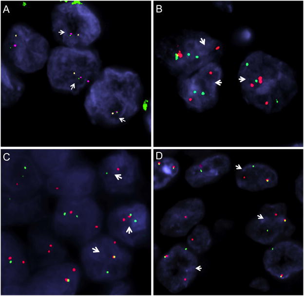
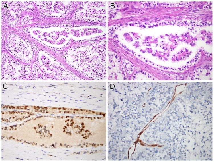
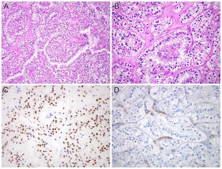
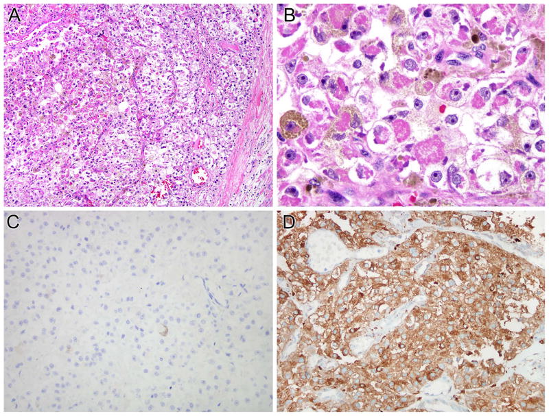
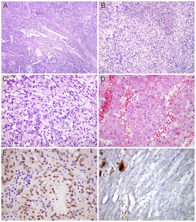
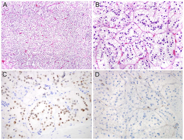
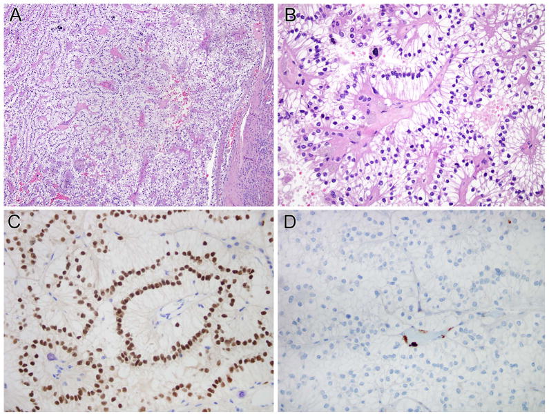
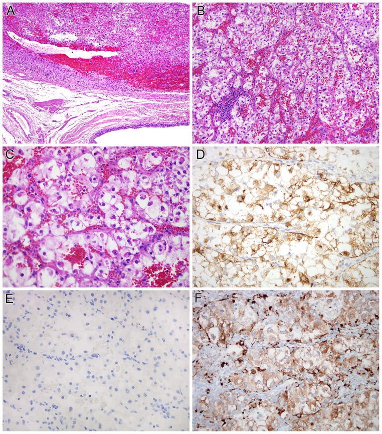
Xp11 translocation cancers include Xp11 translocation renal cell carcinoma (RCC), Xp11 translocation perivascular epithelioid cell tumor (PEComa), and melanotic Xp11 translocation renal cancer. In Xp11 translocation cancers, oncogenic activation of TFE3 is driven by the fusion of TFE3 with a number of different gene partners; however, the impact of individual fusion variant on specific clinicopathologic features of Xp11 translocation cancers has not been well defined. In this study, we analyze 60 Xp11 translocation cancers by fluorescence in situ hybridization using custom bacterial artificial chromosome probes to establish their TFE3 fusion gene partner. In 5 cases RNA sequencing was also used to further characterize the fusion transcripts. The 60 Xp11 translocation cancers included 47 Xp11 translocation RCC, 8 Xp11 translocation PEComas, and 5 melanotic Xp11 translocation renal cancers. A fusion partner was identified in 53/60 (88%) cases, including 18 SFPQ (PSF), 16 PRCC, 12 ASPSCR1 (ASPL), 6 NONO, and 1 DVL2. We provide the first morphologic description of the NONO-TFE3 RCC, which frequently demonstrates subnuclear vacuoles leading to distinctive suprabasal nuclear palisading. Similar subnuclear vacuolization was also characteristic of SFPQ-TFE3 RCC, creating overlapping features with clear cell papillary RCC. We also describe the first RCC with a DVL2-TFE3 gene fusion, in addition to an extrarenal pigmented PEComa with a NONO-TFE3 gene fusion. Furthermore, among neoplasms with the SFPQ-TFE3, NONO-TFE3, DVL2-TFE3, and ASPL-TFE3 gene fusions, the RCCs are almost always PAX8 positive, cathepsin K negative by immunohistochemistry, whereas the mesenchymal counterparts (Xp11 translocation PEComas, melanotic Xp11 translocation renal cancers, and alveolar soft part sarcoma) are PAX8 negative, cathepsin K positive. These findings support the concept that despite an identical gene fusion, the RCCs are distinct from the corresponding mesenchymal neoplasms, perhaps due to the cellular context in which the translocation occurs. We corroborate prior data showing that the PRCC-TFE3 RCCs are the only known Xp11 translocation RCC molecular subtype that are consistently cathepsin K positive. In summary, our data expand further the clinicopathologic features of cancers with specific TFE3 gene fusions and should allow for more meaningful clinicopathologic associations to be drawn.
Figures
Comment in
-
TFE3-Fusion Variant Analysis Defines Specific Clinicopathologic Associations Amog Xp11 Translocation Cancers.Am J Surg Pathol. 2017 Jan;41(1):138-140. doi: 10.1097/PAS.0000000000000730. Am J Surg Pathol. 2017. PMID: 27631518 No abstract available.
References
-
- Argani P, Antonescu CR, Illei PB, Lui MY, Timmons CF, Newbury R, Reuter VE, Garvin AJ, Perez-Atayde AR, Fletcher JA, Beckwith JB, Bridge JA, Ladanyi M. Primary renal neoplasms with the ASPL-TFE3 gene fusion of alveolar soft part sarcoma: A distinctive tumor entity previously included among renal cell carcinomas of children and adolescents. Am J Pathol. 2001;159:179–92. - PMC - PubMed
-
- Argani P, Antonescu CR, Couturier J, Fournet J, Sciot R, Debiec-Rychter M, Hutchinson B, Reuter VE, Boccon-Gibod L, Timmons C, Hafez N, Ladanyi M. PRCC-TFE3 renal carcinomas: Morphologic, immunohistochemical, ultrastructural, and molecular analysis of an entity associated with the t(X;1)(p11. 2;q21) Am J Surg Pathol. 2002;26:1553–66. - PubMed
-
- Clark J, Lu YJ, Sidhar SK, Parker C, Gill S, Smedley D, Hamoudi R, Linehan WM, Shipley J, Cooper CS. Fusion of splicing factor genes PSF and NonO (p54nrb) to the TFE3 gene in papillary renal cell carcinoma. Oncogene. 1997;15:2233–9. - PubMed
-
- Argani P, Lui MY, Couturier J, Bouvier R, Fournet J, Ladanyi M. A novel CLTC-TFE3 gene fusion in pediatric renal adenocarcinoma with t(X;17)(p11. 2;q23) Oncogene. 2003;22:5374–8. - PubMed
Publication types
MeSH terms
Substances
Grants and funding
LinkOut - more resources
Full Text Sources
Other Literature Sources
Medical
Molecular Biology Databases
