Review
. 2016 Jul;40(4):464-79.
doi: 10.1093/femsre/fuw006. Epub 2016 Mar 13.

Bacterial glycobiology: rhamnose-containing cell wall polysaccharides in Gram-positive bacteria

Affiliations
Review

Bacterial glycobiology: rhamnose-containing cell wall polysaccharides in Gram-positive bacteria

Michel-Yves Mistou et al. FEMS Microbiol Rev. 2016 Jul.

Abstract

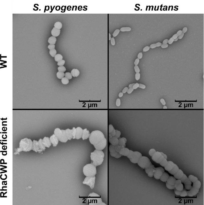
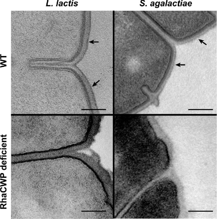
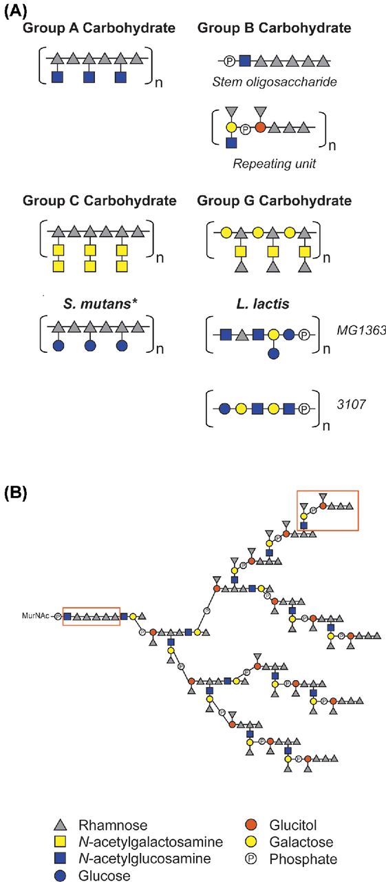
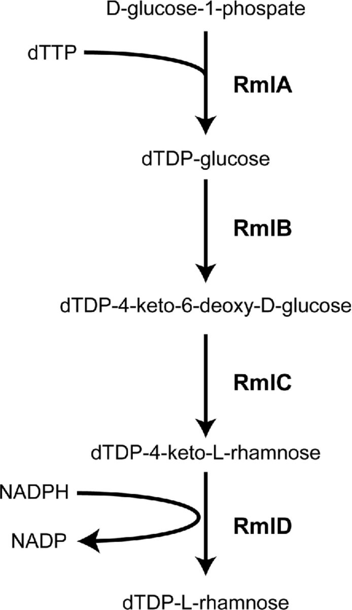
The composition of the Gram-positive cell wall is typically described as containing peptidoglycan, proteins and essential secondary cell wall structures called teichoic acids, which comprise approximately half of the cell wall mass. The cell walls of many species within the genera Streptococcus, Enterococcus and Lactococcus contain large amounts of the sugar rhamnose, which is incorporated in cell wall-anchored polysaccharides (CWP) that possibly function as homologues of well-studied wall teichoic acids (WTA). The presence and chemical structure of many rhamnose-containing cell wall polysaccharides (RhaCWP) has sometimes been known for decades. In contrast to WTA, insight into the biosynthesis and functional role of RhaCWP has been lacking. Recent studies in human streptococcal and enterococcal pathogens have highlighted critical roles for these complex polysaccharides in bacterial cell wall architecture and pathogenesis. In this review, we provide an overview of the RhaCWP with regards to their biosynthesis, genetics and biological function in species most relevant to human health. We also briefly discuss how increased knowledge in this field can provide interesting leads for new therapeutic compounds and improve biotechnological applications.

Keywords: Gram-positive bacteria; biosynthesis; cell wall polysaccharide; glycobiology; pathogenesis; rhamnose.


Figures

Figure 1.
Figure 1.
Cell division and separation defects caused by RhaCWP deficiency in S. mutans and S. pyogenes. Representative scanning electron microscopy images of S. pyogenes and S. mutans wild-type (WT) strains and corresponding RhaCWP-deficient strains. In S. pyogenes loss of the GAC was enforced by inducible knockout of gacA (rmlD homologue) and in S. mutans, deletion of rmlD results in loss of RhaCWP (van der Beek et al. 2015). Scale bar is indicated in image.
Figure 2.
Figure 2.
Visualization of the pellicle by transmission electron microscopy. Lactococcus lactis and S. agalactiae transmission electron microscope images contrasted with heavy metal staining method. In wild-type (WT) strains the pellicle is visible and indicated by arrows. Loss of RhaCWP expression, due to of genetic mutation, result in loss of pellicle expression. The scale bar represents 0.1 μm.
Figure 3.
Figure 3.
Schematic representation of known RhaCWP structures. (A) Schematic representation of RhaCWP core structures in different streptococcal species and L. lactis. GAC structure (McCarty and Lancefield ; Coligan, Kindt and Krause ; Pritchard et al. ; Huang, Rama Krishna and Pritchard 1986); GBC structure (Pritchard, Gray and Dillon ; Michon et al. 1987; 1988); GCC structure (Krause and McCarty ; Coligan, Kindt and Krause 1978); GGC structure (Pritchard et al. 1988); S. mutans serotypes c, e, f, k (Pritchard and Furner ; Pritchard et al. , ; Nakano and Ooshima 2009); L lactis (Chapot-Chartier et al. ; Ainsworth et al. ; Chapot-Chartier and Kulakauskas 2014). *For S. mutans the glucose side chain can either be absent (serotype k) or be linked to the rhamnan backbone in α-1,2 configuration (serotype c), β-1,2 configuration (serotype e), or α-1,3 configuration (serotype f). It must be noted that the (distribution of) length(s) of the RhaCWP have not been experimentally determined. RhaCWP are likely covalently attached to peptidoglycan MurNAc. (B) Full structure of GBC as described by (Pritchard, Gray and Dillon ; Michon et al. 1987, 1988) with newly recognized structural elements highlighted in boxes. Because both the repeating unit and the rhamnan stem have a basal GlcNAc moiety, we hypothesize that the synthesis of each building block is initiated separately on the undecaprenyl lipid carrier by GbcO. It is recognized that either incomplete substitution (at branch points located on the penultimate rhamnose of the repeating unit) or further extension could create a more heterogeneous final structure than presented here. Phosphate groups are involved in phosphodiester bonds linking oligosaccharides into polysaccharides.
Figure 4.
Figure 4.
L-rhamnose biosynthesis pathway. Four-step catalytic reaction resulting in dTDP-L-rhamnose production from D-glucose-1-phosphate.
Figure 5.
Figure 5.
Comparison of the RhaCWP biosynthesis gene clusters in streptococcal species, L. lactis and E. faecalis. The RhaCWP biosynthesis genes are often located in rmlD-associated gene clusters. The loci vary between 14 and 26 kB in size encoding between 12 and 25 genes with annotated functions such as glycosyltransferases, polysaccharide biosynthesis proteins, rhamnose biosynthesis proteins (Rml proteins) and putative transport molecules. Each function is indicated by a different color. A representative gene cluster of a single species is presented and abbreviated gene annotations are indicated below. Arrows are drawn to scale with gene size. The RhaCWP gene clusters of the following strains are shown: S. pyogenes strain M5005 spy0602 – spy0613; S. agalactiae NEM316 gbs1480 – gbs1494; S. equi subs equisimilis MGCS10565 Sez837 – Sez850; S. dysgalactiae sub equisimilis GGS_124 SDEG750 – SDEG764; S. mutans UA159 SMU.824 – SMU.836; E. faecalis V583 EF2198 – EF2174; L. lactis MG1363 llmg0206 – llmg0228. TM, transmembrane
Figure 6.
Figure 6.
General steps in glycoconjugate biosynthesis. Despite glycoconjugate diversity, bacterial glycoconjugate biosynthesis is quite conserved and proceeds in five basic steps: (1) initiation of biosynthesis through activation of a lipid carrier on the cytoplasmic side of the membrane, (2) elongation by sequential addition of activated monosaccharides by glycosyltransferases to form the polysaccharide on the polyprenoid linker, (3) translocation of lipid-linked precursors across the membrane by ABC transporters or ‘flippases’, (4) linkage of the glycoconjugate to peptidoglycan by LytR/CpsA/Psr proteins, and (5) additional modifications to the glycoconjugate that can occur after anchoring to the cell wall, for example alanylation of wall teichoic acid. GlcNAc, N-acetylglucosamine. UDP-GlcNAcP transferase, UDP-GlcNAc:lipid phosphate transferase

References

    1. Adibekian A, Stallforth P, Hecht M-L, et al. Comparative bioinformatics analysis of the mammalian and bacterial glycomes. Chem Sci. 2011;2:337–44.
    1. Ainsworth S, Sadovskaya I, Vinogradov E, et al. Differences in lactococcal cell wall polysaccharide structure are major determining factors in bacteriophage sensitivity. mBio. 2014;5:e00880–14. - PMC - PubMed
    1. Alphey MS, Pirrie L, Torrie LS, et al. Allosteric competitive inhibitors of the glucose-1-phosphate thymidylyltransferase (RmlA) from Pseudomonas aeruginosa. ACS Chem Biol. 2013;8:387–96. - PubMed
    1. Andre G, Kulakauskas S, Chapot-Chartier MP, et al. Imaging the nanoscale organization of peptidoglycan in living Lactococcus lactis cells. Nat Commun. 2010;1:27. - PMC - PubMed
    1. Ayoub EM, Dudding BA. Streptococcal Group A carbohydrate antibody in rheumatic and nonrheumatic bacterial endocarditis. J Lab Clin Med. 1970;76:322–32. - PubMed

MeSH terms