. 2016 May;131(5):725-36.
doi: 10.1007/s00401-016-1555-z. Epub 2016 Mar 14.

Vascular basement membranes as pathways for the passage of fluid into and out of the brain

Affiliations

Vascular basement membranes as pathways for the passage of fluid into and out of the brain

Alan W J Morris et al. Acta Neuropathol. 2016 May.

Abstract

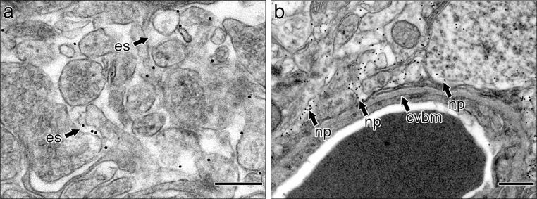
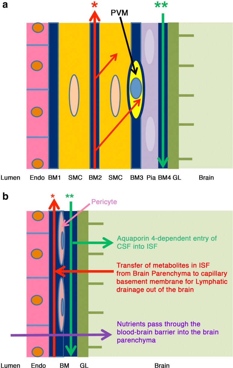
In the absence of conventional lymphatics, drainage of interstitial fluid and solutes from the brain parenchyma to cervical lymph nodes is along basement membranes in the walls of cerebral capillaries and tunica media of arteries. Perivascular pathways are also involved in the entry of CSF into the brain by the convective influx/glymphatic system. The objective of this study is to differentiate the cerebral vascular basement membrane pathways by which fluid passes out of the brain from the pathway by which CSF enters the brain. Experiment 1: 0.5 µl of soluble biotinylated or fluorescent Aβ, or 1 µl 15 nm gold nanoparticles was injected into the mouse hippocampus and their distributions determined at 5 min by transmission electron microscopy. Aβ was distributed within the extracellular spaces of the hippocampus and within basement membranes of capillaries and tunica media of arteries. Nanoparticles did not enter capillary basement membranes from the extracellular spaces. Experiment 2: 2 µl of 15 nm nanoparticles were injected into mouse CSF. Within 5 min, groups of nanoparticles were present in the pial-glial basement membrane on the outer aspect of cortical arteries between the investing layer of pia mater and the glia limitans. The results of this study and previous research suggest that cerebral vascular basement membranes form the pathways by which fluid passes into and out of the brain but that different basement membrane layers are involved. The significance of these findings for neuroimmunology, Alzheimer's disease, drug delivery to the brain and the concept of the Virchow-Robin space are discussed.

Keywords: Alzheimer’s disease; Amyloid-β; Arteries; Basement membranes; Capillaries; Drug delivery; Lymphatic drainage; Nanoparticles neuroimmunology; Virchow–Robin space.


Figures

Fig. 1
Fig. 1
Extracellular space in grey matter of the hippocampus and its connection to capillary basement membrane. a Grey matter with the extracellular space outlined in yellow. There is a capillary in the bottom left-hand corner. Capillary lumen (lu). b High-magnification view of the junction of the extracellular space (es) with the capillary basement membrane (cvbm) at the level of the astrocyte membrane. Endothelial cell of the capillary (en); astrocyte of the glia limitans (a). Scale bars a 500 nm; b 250 nm
Fig. 2
Fig. 2
Biotinylated Aβ identified in the extracellular space of hippocampal grey matter and in a capillary basement membrane. a Hippocampal neuropil showing darkly stained biotinylated Aβ in the extracellular spaces (arrows labelled Aβ); b capillary in the hippocampus showing darkly stained biotinylated Aβ in the basement membrane; c high-magnification view of the area within the square in Fig. 2b showing darkly stained biotinylated Aβ in the extracellular space (right) and throughout the capillary basement membrane (left) that encapsulates a pericyte (p); d capillary from a control hippocampus, into which no biotinylated Aβ has been injected. The basement membrane (cvbm) is negative for Aβ staining. en endothelium, lu lumen, p pericyte, cvbm cerebrovascular basement membrane. Scale bars a 500 nm; bd 200 nm
Fig. 3
Fig. 3
Immunocytochemistry/confocal images showing that 5 min after the injection of soluble Aβ into the hippocampus, Aβ is associated with basement membrane proteins within the wall of an artery in the hippocampal fissure. a Aβ (red) in the wall of a leptomeningeal artery; b laminin (blue); c a layer of smooth muscle cells (green) in the artery wall that is most clearly seen when the vessel is cut in cross section; d a merged image showing co-localisation of Aβ and laminin (purple–see arrows) associated with the smooth muscle cells. The outermost layer of the artery, the tunica adventitia (ta), is stained only for laminin (blue) and not for Aβ. SP5 Leica confocal image. Scale bar 50 μm
Fig. 4
Fig. 4
Nanoparticles are distributed in the extracellular spaces but do not enter the capillary basement membrane. a Densely stained punctate nanoparticles are seen in the extracellular spaces (es) of the brain and within cell processes of neurons and possibly of astrocytes. b Neuropil and capillary showing nanoparticles (np) in the extracellular spaces of the neuropil and occasionally in cell processes but no nanoparticles are located in the capillary basement membrane (cvbm). Scale bars a 300 nm; b 500 nm
Fig. 5
Fig. 5
Distribution in the brain of 15 nm nanoparticles injected into the CSF. Groups of 15 nm nanoparticles are present in the pial-glial basement membrane of a cortical artery 5 min after injection into the CSF of the cisterna magna. a Low-power transmission electronmicrograph of an artery near the surface of the cerebral cortex. Note that cells and basement membranes form compact layers and there is no Virchow–Robin space. Dense nanoparticles form lines or groups in the pial-glial basement membrane between the pia mater and the glia limitans around nearly half the circumference of the artery but no nanoparticles are seen in basement membranes between smooth muscle cells. b Higher magnification of the square labelled 1 in a. Layers of the artery wall can be identified moving outwards from the lumen (lu) through the endothelium (en), smooth muscle coat (sm) and a layer of pia mater (pm) to the glia limitans of the brain parenchyma (pa). Dense groups and single nanoparticles (np) are located mainly in the pial-glial basement membrane (cvbm) between the pia mater (pm) and the adjacent glia limitans. Scale bars a 1 μm; b 500 nm
Fig. 6
Fig. 6
Schematic representation of the lymphatic drainage and convective influx/glymphatic systems of the brain. An artery enters the brain from the subarachnoid space and an arteriole divides into capillaries. At the top of the figure, the artery is lined by endothelium (Endo), and coated by the tunica media (TM) composed of smooth muscle cells and by the outermost tunica adventitia (TA) composed of connective tissue. As it enters the brain, the artery loses the tunica adventitia but is still coated by a layer of pia-arachnoid (Pia) that intervenes between the artery and the glia limitans (GL) of the brain. As the arteriole divides into capillaries, the tunica media and the layer of pia mater are lost. Thus, at the level of the capillary, the glia limitans is in direct contact with the wall of the capillary. On the right-hand side of the diagram, the red arrows indicate the intramural perivascular lymphatic drainage pathway by which interstitial fluid (ISF) and solutes pass out of the brain along basement membranes in the walls of capillaries and along basement membranes surrounding smooth muscle cells in the tunica media of arterioles and arteries [7]. Tracers in the CSF enter the brain along the pial-glial basement membrane between the pia mater and the glia limitans (indicated by a green arrow) and enter the brain parenchyma and interstitial fluid by an aquaporin 4-dependent mechanism, which is the convective influx/glymphatic pathway
Fig. 7
Fig. 7
Expanded view of the wall of a cerebral artery near the surface of the cerebral cortex. a The lymphatic drainage pathway for interstitial fluid (ISF) and solutes is specifically along basement membranes surrounding smooth muscle cells in the tunica media (red arrow *). Tracer studies have shown that solutes are taken up by smooth muscle cells and by perivascular macrophages (PVM) (thin red arrows). The convective influx/glymphatic pathway by which CSF enters the brain is along the pial-glial basement membrane (BM4–green arrow **) between the layer of pia mater and the glia limitans of the brain parenchyma. Abbreviations for structure of the artery wall: Endo endothelium, BM basement membrane, SMC smooth muscle cells, GL glia limitans, BM1 basement membrane formed by the endothelium and adjacent smooth muscle cells, BM2 basement membrane formed by adjacent smooth muscle cells; this is the pathway for the lymphatic drainage of interstitial fluid and solutes from the brain along the tunica media, BM3 basement membrane formed by the outer smooth muscle cells and pia mater cells coating the artery, BM4 basement membrane formed by the pia mater and astrocytes of the glia limitans. It is along BM4 that CSF and nanoparticles enter the brain in the convective influx/glymphatic system. b Expanded view of the wall of a cerebral capillary and surrounding glia limitans. The endothelium has a basement membrane that is formed partly by endothelial cells and partly by astrocytes of the glia limitans. Solutes, such as amyloid β (Aβ) in the interstitial fluid pass from the extracellular spaces of the brain into the endothelial-glial basement membrane and drain along the intramural perivascular drainage pathway (red arrow). Tracers injected into the CSF reach the capillary basement membranes via the convective influx/glymphatic system and their entry into the interstitial fluid is dependent upon aquaporin 4 (green arrows). Nutrients from the blood through the blood–brain barrier pass into the brain through the endothelium, the basement membrane and the glia limitans (purple arrow). Pericytes are surrounded by basement membrane and lie between the endothelium (Endo) and the basement membrane (BM) of the glia limitans (GL). Although the red and green arrows are shown in separate parts of the basement membrane, a single capillary basement membrane appears to share traffic of fluid into and out of the brain

References

    1. Abbott NJ. Dynamics of CNS barriers: evolution, differentiation, and modulation. Cell Mol Neurobiol. 2005;25:5–23. doi: 10.1007/s10571-004-1374-y. - DOI - PMC - PubMed
    1. Abbott NJ. Evidence for bulk flow of brain interstitial fluid: significance for physiology and pathology 1. Neuro Chem Int. 2004;45:545–552. doi: 10.1016/j.neuint.2003.11.006. - DOI - PubMed
    1. Barua NU, Bienemann AS, Hesketh S, Wyatt MJ, Castrique E, Love S, Gill SS. Intrastriatal convection-enhanced delivery results in widespread perivascular distribution in a pre-clinical model. Fluids Barriers CNS. 2012;9:2. doi: 10.1186/2045-8118-9-2. - DOI - PMC - PubMed
    1. Bedussi B, van Lier MG, Bartstra JW, de Vos J, Siebes M, VanBavel E, Bakker EN. Clearance from the mouse brain by convection of interstitial fluid towards the ventricular system. Fluids Barriers CNS. 2015;12:23. doi: 10.1186/s12987-015-0019-5. - DOI - PMC - PubMed
    1. Brinker T, Stopa E, Morrison J, Klinge P. A new look at cerebrospinal fluid circulation. Fluids Barriers CNS. 2014;11:10. doi: 10.1186/2045-8118-11-10. - DOI - PMC - PubMed

Publication types

MeSH terms