. 2016 May 6;291(19):10006-20.
doi: 10.1074/jbc.M116.717405. Epub 2016 Mar 14.

Mechanism of Concerted RNA-DNA Primer Synthesis by the Human Primosome

Affiliations

Mechanism of Concerted RNA-DNA Primer Synthesis by the Human Primosome

Andrey G Baranovskiy et al. J Biol Chem. .

Abstract

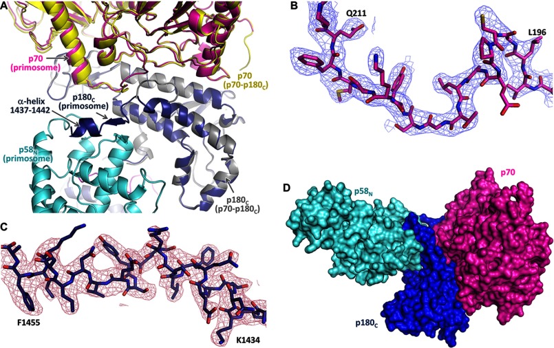
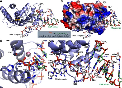
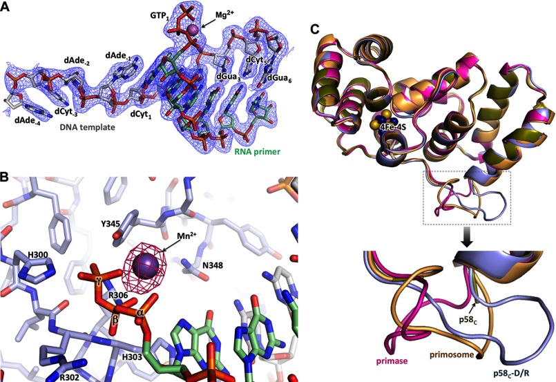
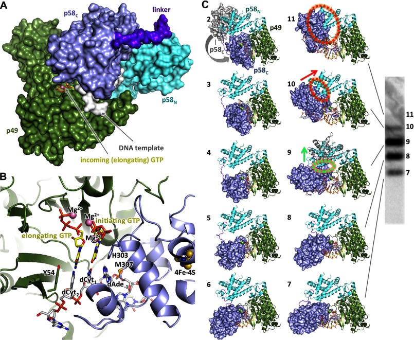
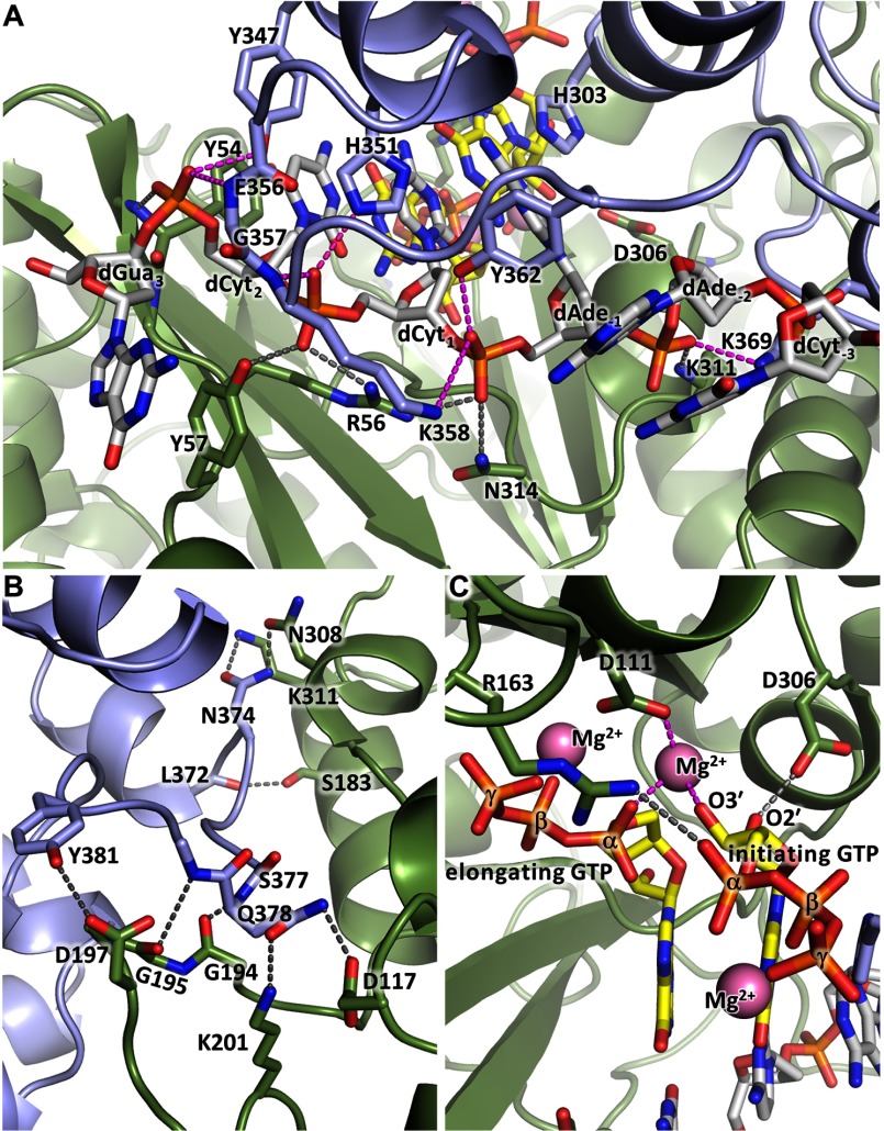
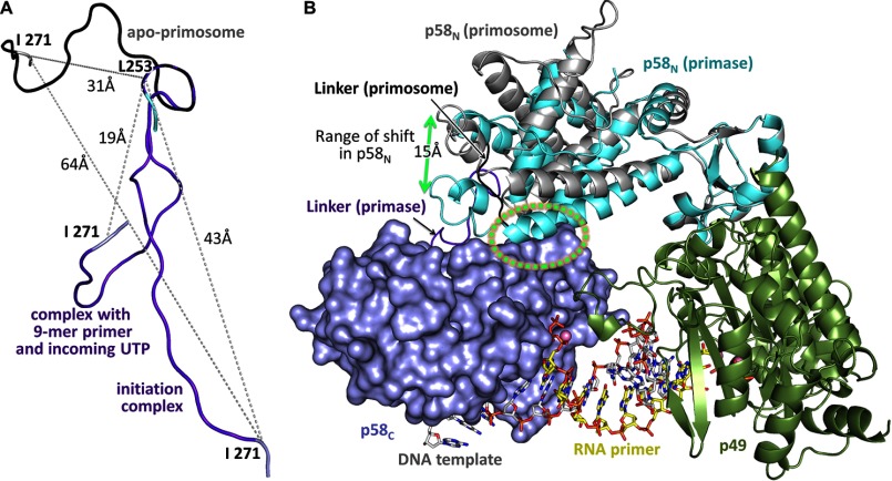
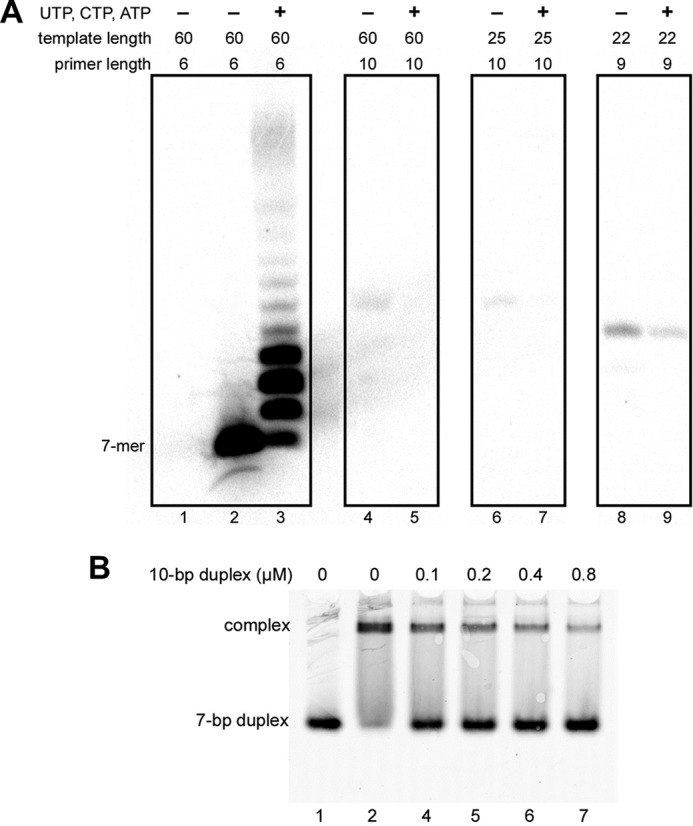
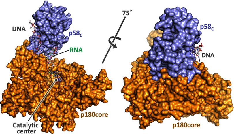
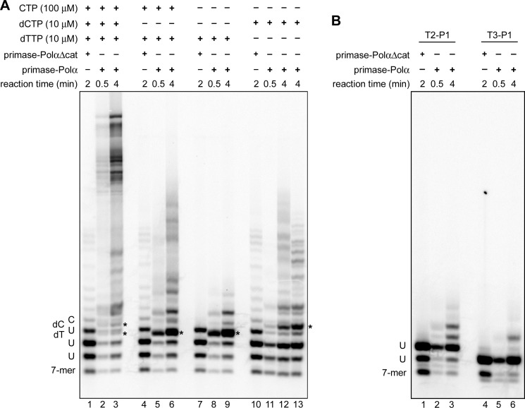
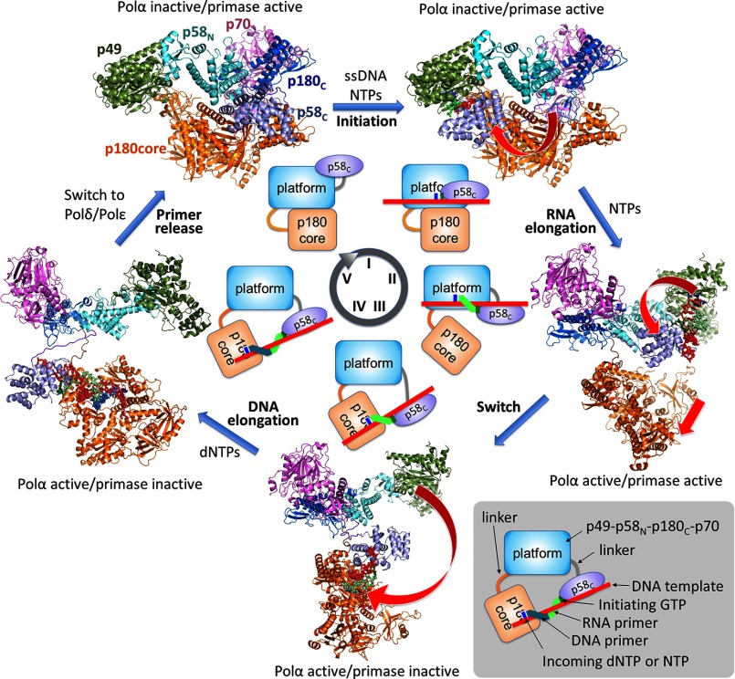
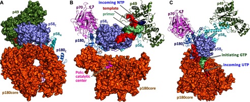
The human primosome, a 340-kilodalton complex of primase and DNA polymerase α (Polα), synthesizes chimeric RNA-DNA primers to be extended by replicative DNA polymerases δ and ϵ. The intricate mechanism of concerted primer synthesis by two catalytic centers was an enigma for over three decades. Here we report the crystal structures of two key complexes, the human primosome and the C-terminal domain of the primase large subunit (p58C) with bound DNA/RNA duplex. These structures, along with analysis of primase/polymerase activities, provide a plausible mechanism for all transactions of the primosome including initiation, elongation, accurate counting of RNA primer length, primer transfer to Polα, and concerted autoregulation of alternate activation/inhibition of the catalytic centers. Our findings reveal a central role of p58C in the coordinated actions of two catalytic domains in the primosome and ultimately could impact the design of anticancer drugs.

Keywords: 5′-triphosphate; DNA polymerase; DNA primase; DNA-protein interaction; DNA/RNA duplex; RNA primer length counting; RNA synthesis; human DNA replication; initiating NTP; x-ray crystallography.


Figures

FIGURE 1.
FIGURE 1.
Crystallization of the primosome and p58C-D/R. A and B, photomicrographs of the primosome crystals before and after optimization, respectively. C, a single crystal of primosome scooped in a nylon-fiber loop. D, analysis of the content of primosome crystals by 7% SDS-PAGE. Lane 1, molecular weight markers; lanes 2 and 3 correspond to the crystals shown in A and B, respectively. E and F, photomicrographs of p58C-D/R crystals in the form of plates and needles, respectively. G, analysis of the content of a needle-like p58C-D/R crystal by 5% native PAGE. Gel was stained first with ethidium bromide (left panel) and then with Coomassie R-250 (right panel).
FIGURE 2.
FIGURE 2.
Structure of the human primosome hetero-tetramer complex. A, schematic representation of the domain organization. Flexibly tethered domains are shown as separate parts. p58C coordinates the iron-sulfur cluster. Exo* is an exonuclease domain with no associated activity due to evolutionary substitution of the catalytic amino acid residues; alignment is as described in Ref. . PDE, phosphodiesterase. B, the crystal structure of primosome. Subunits are shown as schematics and colored as in A. The α-carbons of catalytic aspartates are shown as purple spheres.
FIGURE 3.
FIGURE 3.
Mechanism of Polα autoinhibition in non-polymerizing primosome. A, Polα structure in apo-primosome. Zn2 module of p180C and OB domain of p70 block the entry to the Polα active site for a template/primer. PDE, phosphodiesterase. B, alignment of Polα catalytic cores in the ternary complex with substrates (colored gray; PDB code 4QCL) and in apo-primosome (colored as in A) shows a significant difference in positions of the palm and thumb relative to other subdomains.
FIGURE 4.
FIGURE 4.
Domain and linkers organization in the primosome. A, primosome has two long linkers connecting p180core and p58C to the platform composed of p49-p58N-p180C-p70. Primase linker (colored red) connects p58C and p58N; Polα linker (colored green) connects p180core and p180C. B, analysis of the electron density map for p180 residues 1243–1250 confirms their packing as α-helix. Nitrogens, oxygens, and carbons are colored blue, red, and orange, respectively. C, the carbons in p58N, p58C, and the linker are colored cyan, gray, and magenta, respectively. D, the relative positions of p58C in the primase (PDB ID 4RR2) and the primosome structures (only p58N was used for alignment).
FIGURE 5.
FIGURE 5.
The regions disordered in Polα are structured in primosome. A, superimposition of p180C-p70 (PDB code 4Y97) and primosome structures. In primosome, p180C and p70 are colored magenta and blue; in p180C-p70, they are colored yellow and gray. B and C, 2FoFc Fourier map (contour level at 1σ) for p70 residues 195–212 and p180C residues 1433–1456, respectively. D, the relative position of p70 and p58N is stabilized by p180C.
FIGURE 6.
FIGURE 6.
Structure of p58C in complex with the RNA-primed DNA template. A, specific recognition of the template/primer junction at the 5′ terminus of the primer by p58C. B, the surface electrostatic potential of p58C shows docking of the template and the 5′-triphosphate of the primer on two separate positively charged areas (colored blue). C, hydrogen bonds (dashed lines) between p58C and the β- and γ-phosphates of GTP1. D, hydrogen bonds between p58C and the template.
FIGURE 7.
FIGURE 7.
Details of p58C interaction with a template/primer. A, electron density map for the DNA/RNA duplex and the triphosphate coordinating Mg2+. The carbons of DNA and RNA are colored green and gray, respectively. B, FoFc Fourier map (contour level at 5σ) for Mn2+ coordinated by a 5′-triphosphate of RNA. C, alignment of p58C from different structures points to the flexibility of the DNA-interacting loop 354–366. The PDB accession numbers for the structures of the human primase and p58C are 4RR2 and 3Q36, respectively.
FIGURE 8.
FIGURE 8.
Mechanism of RNA synthesis initiation, elongation, and termination. A, compact shape of primase in the initiation complex model. B, close-up view of the initiation mini duplex in the model. Position of the magnesium ion between the elongating and initiating GTPs was modeled to coordinate Asp-111, α-phosphate of elongating GTP, and O3′ of initiating GTP as in the active site of polymerases (58). C, models of the initiation and elongation complexes. The crystal structures of p58C-D/R, human primase (PDB code 4RR2), and p49-p58N–UTP complex (PDB code 4BPW) were used to build these models. Panel numbers correspond to the length of the growing RNA primer plus incoming NTP in the modeled complexes. Panel 2 corresponds to the initiation complex resulting in dinucleotide formation and also shows the position of p58C in apo-primase. The clash area is depicted by dotted circles (colored green or red to indicate avoidable or unavoidable steric hindrance, respectively), whereas the arrows show the direction at which p58C is pushing p58N. Close-up view of the complex in panel 9, explaining how steric hindrance is avoided, is provided in Fig. 10B. The image on the right shows the typical pattern of primase-catalyzed reaction products.
FIGURE 9.
FIGURE 9.
p49-DNA and p49-p58C interaction interfaces in the model of initiation complex. A, interaction of the DNA template with p49 and p58C. The hydrogen bonds between DNA and p58C or p49 are depicted by pink or gray dashed lines, respectively. B, the potential p49-p58C interaction interface shows its plasticity due to the absence of hydrophobic contacts. C, p49 interacts with a ribose and α-phosphate of the initiating GTP. Second magnesium ion was modeled as described in the legend to Fig. 8B.
FIGURE 10.
FIGURE 10.
Conformational changes in human primase during RNA primer synthesis. A, position of the p58N-p58C linker in apo-primosome (Fig. 2B), in the initiation complex (Fig. 8A) and in the primase complex with a DNA template primed by a 9-mer primer and the incoming UTP (Fig. 8C, panel 10). The alignment is performed using p58N. B, close-up view of the primase-DNA/RNA-CTP elongation complexes containing an 8-mer primer. The complexes were modeled based on the structures of primosome (p58N is colored gray), primase (p58N is colored cyan; PDB code 4RR2), and p58C-D/R. The alignment was performed using p49 subunits. In the primosome, only p58N with the p58N-p58C linker is shown for clarity. Clash area between the two domains in the primase-based model is depicted by the green dotted circle; no clash is observed in the primosome-based model.
FIGURE 11.
FIGURE 11.
DNA/RNA duplex length affects primase activity but not affinity. A, the effect of RNA primer length on activity of primase-PolαΔcat. Lanes 1–3, T1-P1; lanes 4 and 5, T6-P4; lanes 6 and 7, T7-P4; lanes 8 and 9, T5-P3. Reactions corresponding to lanes 2, 4, 6, and 8 were conducted in the presence of [α-33P]GTP alone to show the position of the first product of primer extension. Reactions corresponding to lanes 3, 5, 7, and 9 were supplemented with 0.1 mm each UTP, CTP, and ATP. Reactions were run for 2 min at 35 °C, and products were resolved by 20% urea-PAGE. B, 10-bp DNA/RNA duplex (T7-P4) efficiently competes with a 7-bp duplex (T4-P2) for binding with primase-PolαΔcat. All reactions contained 0.2 μm T4-P2; reactions corresponding to lanes 2–7 contained 0.2 μm primase-PolαΔcat. Samples were resolved by 5% native PAGE and visualized using the Typhoon 9410 imager. T4 is labeled with Cy3 at the 5′-end.
FIGURE 12.
FIGURE 12.
The model of the switch complex. The crystal structures of p58C-D/R and p180core-D/R-dCTP (PDB code 4QCL) were used for model building.
FIGURE 13.
FIGURE 13.
Determination of the Polα start position during RNA primer extension with primosome. A and B, analysis of the ribo- and deoxy-NTPs incorporation using the 6-mer RNA primer P1 annealed to the 60-mer DNA template T1 (A) or to the 59-mer and 58-mer DNA templates T2 and T3, respectively (B). All reactions with the T1-P1 duplex contained 100 μm UTP; all reactions with T2-P1 and T3-P1 duplexes contained 100 μm UTP and 10 μm dTTP. Primase-PolαΔcat is the primosome without p180core.
FIGURE 14.
FIGURE 14.
Mechanism of RNA-DNA primer synthesis by the human primosome. At the first step (steps are labeled by roman numerals at the circle in the center), p58C moves toward p49 (curved arrow) to initiate RNA synthesis. During the second step, p58C is moving toward p180core by following the helical path of the growing DNA/RNA duplex (curved arrow) and pushes the p180core to dissociate from the platform (straight arrow). Additionally, when RNA primer length is nine nucleotides, p58C makes a steric hindrance with p58N, which prevents primer extension by p49. At the third step, p58C rotates (curved arrow) and loads the template/primer to the Polα active site. At the fourth step, Polα extends the RNA primer with dNTPs, whereas p58C is holding the 5′ terminus of the primer. At the fifth step, primosome is replaced by Polϵ or Polδ. Models of primosome at different steps of primer synthesis are based on the apo-primosome structure (Fig. 2) and primase models (Fig. 8).
FIGURE 15.
FIGURE 15.
Models of the human primosome during key steps of RNA primer synthesis. A, at the initiation step, p58C fits into a space between p180core and p49. B, at the RNA elongation step, p58C is moving toward p180core and displaces it from the p49-p58N-p180C-p70 platform. C, flexibly tethered p180core binds DNA template with a 9-mer RNA primer presented by p58C.

References

    1. Pellegrini L. (2012) The Pol α-primase complex. Subcell. Biochem. 62, 157–169 - PubMed
    1. Muzi-Falconi M., Giannattasio M., Foiani M., and Plevani P. (2003) The DNA polymerase α-primase complex: multiple functions and interactions. ScientificWorldJournal 3, 21–33 - PMC - PubMed
    1. Yeeles J. T., Deegan T. D., Janska A., Early A., and Diffley J. F. (2015) Regulated eukaryotic DNA replication origin firing with purified proteins. Nature 519, 431–435 - PMC - PubMed
    1. Reijns M. A., Kemp H., Ding J., de Procé S. M., Jackson A. P., and Taylor M. S. (2015) Lagging-strand replication shapes the mutational landscape of the genome. Nature 518, 502–506 - PMC - PubMed
    1. Pavlov Y. I., Frahm C., Nick McElhinny S. A., Niimi A., Suzuki M., and Kunkel T. A. (2006) Evidence that errors made by DNA polymerase α are corrected by DNA polymerase δ. Curr. Biol. 16, 202–207 - PubMed

Publication types

MeSH terms