

Survival in Mesenchymal Chondrosarcoma Varies Based on Age and Tumor Location: A Survival Analysis of the SEER Database
- PMID: 26975384
- PMCID: PMC5289165
- DOI: 10.1007/s11999-016-4779-2
Survival in Mesenchymal Chondrosarcoma Varies Based on Age and Tumor Location: A Survival Analysis of the SEER Database
Abstract
Background: Studies suggest that mesenchymal chondrosarcoma is associated with a poorer prognosis and a higher proportion of extraskeletal tumors than conventional chondrosarcoma. However, these investigations have been small heterogeneous cohorts, limiting analysis of prognostic factors.
Questions/purposes: (1) What is the 5- and 10-year survival rate of patients diagnosed with mesenchymal chondrosarcoma? (2) What is the effect of demographic and tumor characteristics on survival in patients with mesenchymal chondrosarcoma?
Methods: The Surveillance, Epidemiology, and End Results (SEER) database was used to identify all patients diagnosed with mesenchymal chondrosarcoma from 1973 to 2011. SEER reports survival data on over 8.2 million patients with cancer and has attained 98% completeness in reporting. Using variables within the database, this study designated each patient's tumor as skeletal or extraskeletal and cranial, axial, or appendicular, respectively. Overall survival (OS) was determined for the entire series as well as each group. Median survival was calculated using Kaplan-Meier methods. Cox proportional hazards regression was used to determine whether demographic and tumor variables affected survival. Two hundred five patients with mesenchymal chondrosarcoma were identified, including 82 (40%) skeletal and 123 (60%) extraskeletal.
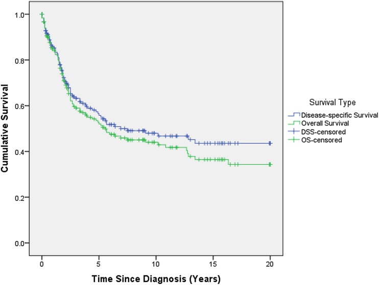
Results: OS for the entire series was 51% (95% confidence interval [CI], 43%-58%) and 43% (95% CI, 35%-51%) at 5 and 10 years, respectively. No difference in OS was detected between extraskeletal and skeletal tumors. Kaplan-Meier analyses showed OS was worse for tumors in axial locations compared with appendicular and cranial locations. Appendicular tumors demonstrated an OS of 50% (95% CI, 36%-63%) at 5 years and 39% (95% CI, 26%-52%) at 10 years. OS for axial tumors was 37% (95% CI, 25%-49%) and 31% (95% CI, 20%-43%), whereas it was 74% (95% CI, 59%-84%) and 67% (95% CI, 50%-79%) for cranial tumors at 5 and 10 years, respectively. When controlling for age, sex, tumor origin, and tumor location, the presence of metastasis (hazard ratio [HR], 12.38; 95% CI, 5.75-26.65; p < 0.001) and 1-cm size increase (HR, 1.16; 95% CI, 1.09-1.23; p < 0.001) were both independently associated with an increased risk of death. Tumor location showed different behaviors depending on patient age. In comparison to cranial tumors at age 20 years, the HR was 5.56 (95% CI, 1.47-21.05; p = 0.01) for axial tumors and 6.26 (95% CI, 1.54-25.42; p = 0.01) for appendicular tumors. At age 60 years, those ratios were 0.10 (95% CI, 0.02-0.55; p = 0.01) and 0.14 (95% CI, 0.04-0.58; p = 0.01), respectively.
Conclusions: Our data suggest that extraskeletal tumors are more common than previously reported; however, this factor does not have clear prognostic value. Presence of metastatic disease and increased tumor size are the main predictors of poor survival outcome. Cranial tumors appear to have a different clinical behavior with our data suggesting better overall survival in young patients (compared with axial and appendicular locations) and a worse survival outcome in older patients.
Level of evidence: Level IV, prognostic study.
Figures
Comment in
-
CORR Insights®: Survival in Mesenchymal Chondrosarcoma Varies Based on Age and Tumor Location: A Survival Analysis of the SEER Database.Clin Orthop Relat Res. 2017 Mar;475(3):806-807. doi: 10.1007/s11999-016-4818-z. Epub 2016 Apr 5. Clin Orthop Relat Res. 2017. PMID: 27048221 Free PMC article. No abstract available.
References
-
- Cesari M, Bertoni F, Bacchini P, Mercuri M, Palmerini E, Ferrari S. Mesenchymal chondrosarcoma. An analysis of patients treated at a single institution. Tumori. 2007;93:423–427. - PubMed
-
- Edge S, Byrd DR, Compton CC, Fritz AG, Greene FL, Trotti A, editors. AJCC Cancer Staging Manual. 7. New York, NY, USA: Springer-Verlag; 2010.
Publication types
MeSH terms
LinkOut - more resources
Full Text Sources
Other Literature Sources
Medical
Research Materials
