. 2016 Apr 19;49(4):695-702.
doi: 10.1021/acs.accounts.5b00538. Epub 2016 Mar 15.

Metalloprotein Crystallography: More than a Structure

Affiliations

Metalloprotein Crystallography: More than a Structure

Sarah E J Bowman et al. Acc Chem Res. .

Abstract

Metal ions and metallocofactors play important roles in a broad range of biochemical reactions. Accordingly, it has been estimated that as much as 25-50% of the proteome uses transition metal ions to carry out a variety of essential functions. The metal ions incorporated within metalloproteins fulfill functional roles based on chemical properties, the diversity of which arises as transition metals can adopt different redox states and geometries, dictated by the identity of the metal and the protein environment. The coupling of a metal ion with an organic framework in metallocofactors, such as heme and cobalamin, further expands the chemical functionality of metals in biology. The three-dimensional visualization of metal ions and complex metallocofactors within a protein scaffold is often a starting point for enzymology, highlighting the importance of structural characterization of metalloproteins. Metalloprotein crystallography, however, presents a number of implicit challenges including correctly incorporating the relevant metal or metallocofactor, maintaining the proper environment for the protein to be purified and crystallized (including providing anaerobic, cold, or aphotic environments), and being mindful of the possibility of X-ray induced damage to the proteins or incorporated metal ions. Nevertheless, the incorporated metals or metallocofactors also present unique advantages in metalloprotein crystallography. The significant resonance that metals undergo with X-ray photons at wavelengths used for protein crystallography and the rich electronic properties of metals, which provide intense and spectroscopically unique signatures, allow a metalloprotein crystallographer to use anomalous dispersion to determine phases for structure solution and to use simultaneous or parallel spectroscopic techniques on single crystals. These properties, coupled with the improved brightness of beamlines, the ability to tune the wavelength of the X-ray beam, the availability of advanced detectors, and the incorporation of spectroscopic equipment at a number of synchrotron beamlines, have yielded exciting developments in metalloprotein structure determination. Here we will present results on the advantageous uses of metals in metalloprotein crystallography, including using metallocofactors to obtain phasing information, using K-edge X-ray absorption spectroscopy to identify metals coordinated in metalloprotein crystals, and using UV-vis spectroscopy on crystals to probe the enzymatic activity of the crystallized protein.


Conflict of interest statement

The authors declare no competing financial interest.

Figures

Figure 1
Figure 1
Anaerobic and aphotic crystallization setups. (A) Setup for metalloprotein crystallization under anaerobic conditions in an MBraun glovebox. Screens can be set up on the mosquito robot; crystal trays are stored and automatically imaged in the Formulatrix RI-182 within a custom designed chamber (Rebekah Bjork, Drennan Laboratory). (B) Light sensitive crystals are imaged and looped in a dark room equipped with red light.
Figure 2
Figure 2
The X-ray beam causes reduction of the bound metallocofactor highlighting the importance of parallel techniques in metalloprotein crystallography. Characteristic bands indicate a low-spin Fe3+ state prior to X-ray exposure; appearance of the 613 nm band postexposure suggests some high-spin Fe2+, consistent with ligand loss. In the NeN64D variant, low-spin Fe3+ is reduced to low-spin Fe2+, evidenced by Q-bands at 520 and 550 nm. Reproduced with permission from ref (44). Copyright 2013 Wiley-VCH Verlaf GmbH&Co, KGaA, Weinheim.
Figure 3
Figure 3
Crystal structure of SyrB2 provides insight into the halogenation mechanism. (A.) Active site of SyrB2, detailing an octahedral iron (brown sphere) geometry with two histidine ligands, α-ketoglutarate, water (blue sphere), and a chloride ion (green sphere). 2FoFc electron density maps are shown in blue mesh and contoured at 1.0σ. (B.) A dispersive difference Fourier map from a SyrB2 crystal that contained bromide (purple sphere). This map, calculated by subtracting data collected at the iron edge (1.7340 Å) from data collected at the bromide edge (0.9197 Å), shows a positive density peak for bromine (purple mesh contoured at 4.0σ), and a negative density peak for iron (brown mesh contoured at −4.0σ) due to the differential scattering of these two ions at these wavelengths. Panel B was reprinted from ref (37).
Figure 4
Figure 4
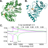
Data collected at the Cu Kα edge can be used to determine phases for Fe–S cluster containing protein structures. The crystal structures of (A) anSME and (B) QueE were solved using the anomalous signal of protein-bound [4Fe–4S] clusters and data collected at the Cu Kα edge. (C) Calculated anomalous scattering at the Fe and Cu K edge. Plot generated with edgeplots web tool from http://skuld.bmsc.washington.edu/scatter/.
Figure 5
Figure 5
Bound metallocofactor AdoCbl is exploited for phase information in CarH. The crystal structure of two domains of the AdoCbl-dependent transcription factor, CarH, was solved using anomalous scattering of the cobalt ion. (A) Experimental electron density maps contoured at 1.2σ (purple) and anomalous electron density map contoured at 5.0σ (yellow). (B) Ribbon trace of AdoCbl, the Cbl-binding and helix bundle domains built into the electron density. (C) Resulting structure of the C-terminal light sensing domains of CarH.
Figure 6
Figure 6
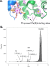
A number of techniques can be used to identify metals within metalloprotein crystals. (A) Anomalous maps (1.54178 Å) contoured at 2.0σ are consistent with Mn2+ in the Mn2+ site and Ca2+ in only one of the two expected calcium-binding sites shown in CP. (B) EDX spectrum of a Mn(II)Ca(II)-bound CP indicates presence of both metals (* indicates presence of K+). Panel A was reprinted from ref (57).
Figure 7
Figure 7
Parallel UV–vis spectroscopy and X-ray crystallography lend support to an ordered mechanism proposed for AxNiR. The intense peak at 595 nm is characteristic of a type I Cu2+ site and disappears after exposure to X-rays, consistent with reduction. Reproduced with permission from ref (18). Copyright 2008 Elsevier Ltd.
Figure 8
Figure 8
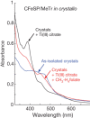
UV–vis of the CFeSP/MeTr crystals show that crystalline protein is active. The spectrum of the CFeSP/MeTr (as-isolated) crystal has features at ∼400 and 470 nm, corresponding to the [4Fe–4S] cluster and Cbl cofactor. Reducing the crystal with Ti(III)-citrate yields a sharp peak at 390 nm, consistent with active Co(I)–Cbl. Addition of CH3–H4folate results in a decrease at 390 nm and a new peak at 450 nm, characteristic of product complex for protein-bound CH3–Co(III). Figure is reprinted from ref (15).

References

    1. Zheng H.; Chruszcz M.; Lasota P.; Lebioda L.; Minor W. Data mining of metal ion environments present in protein structures. J. Inorg. Biochem. 2008, 102, 1765–1776. 10.1016/j.jinorgbio.2008.05.006. - DOI - PMC - PubMed
    1. Waldron K. J.; Rutherford J. C.; Ford D.; Robinson N. J. Metalloproteins and metal sensing. Nature 2009, 460, 823–830. 10.1038/nature08300. - DOI - PubMed
    1. Lippard S. J.; Berg J. M.. Principles of Bioinorganic Chemistry; University Science Books: Mill Valley, CA, 1994.
    1. Rosenzweig A. C. Metallochaperones: bind and deliver. Chem. Biol. 2002, 9, 673–677. 10.1016/S1074-5521(02)00156-4. - DOI - PubMed
    1. Zheng L.; Cash V. L.; Flint D. H.; Dean D. R. Assembly of iron-sulfur clusters. Identification of an iscSUA-hscBA-fdx gene cluster from Azotobacter vinelandii. J. Biol. Chem. 1998, 273, 13264–13272. 10.1074/jbc.273.21.13264. - DOI - PubMed

Publication types

MeSH terms

Substances

LinkOut - more resources