Case Reports
. 2016 May;6(5):492-500.
doi: 10.1158/2159-8290.CD-15-1335. Epub 2016 Mar 14.

Clinical Response of Carcinomas Harboring the BRD4-NUT Oncoprotein to the Targeted Bromodomain Inhibitor OTX015/MK-8628

Affiliations
Case Reports

Clinical Response of Carcinomas Harboring the BRD4-NUT Oncoprotein to the Targeted Bromodomain Inhibitor OTX015/MK-8628

Anastasios Stathis et al. Cancer Discov. 2016 May.

Abstract

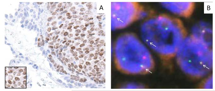
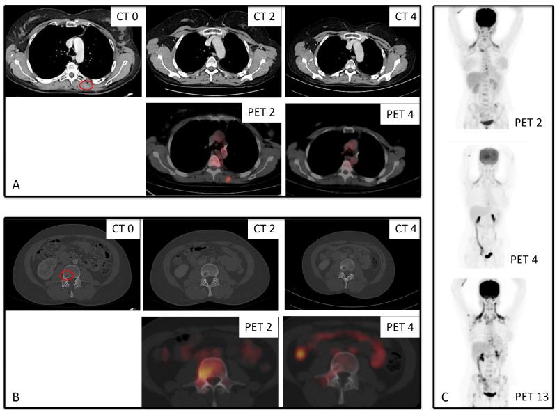
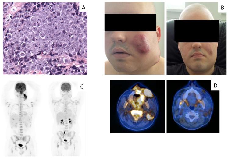
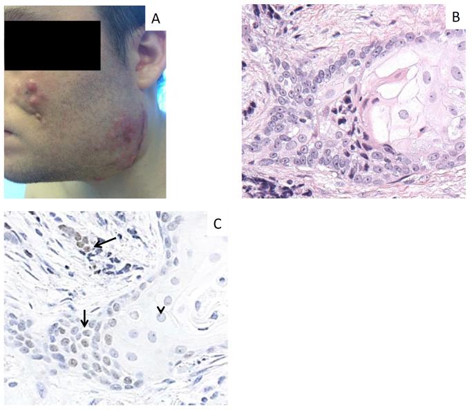
The antineoplastic, prodifferentiative effects of bromodomain and extra-terminal (BET) bromodomain (BRD) inhibitors were initially discovered in NUT midline carcinoma (NMC), an aggressive subtype of squamous cancer driven by the BRD4-NUT fusion oncoprotein. BRD4-NUT blocks differentiation and maintains tumor growth through a potent chromatin-modifying mechanism. OTX015/MK-8628, a novel oral BET inhibitor, targets BRD2/3/4/T with preclinical activity in NMC and several other tumor types and is currently in clinical development. Antitumor activity was evaluated in four patients with advanced-stage NMC with confirmed BRD4-NUT fusions who were treated with 80 mg OTX015/MK-8628 once daily in a compassionate-use context. Two patients responded rapidly with tumor regression and symptomatic relief, and a third had meaningful disease stabilization with a minor metabolic response. The main side effects were mild to moderate gastrointestinal toxicity and fatigue, and reversible grade 3 thrombocytopenia. This is the first proof-of-concept evidence of clinical activity of a BRD inhibitor in targeting BRD4-NUT.

Significance: We present the first clinical proof-of-concept that targeting BRD4-NUT with a BET inhibitor results in impressive and rapid antitumor activity in NMC. It offers strong potential for future clinical application in this rare patient population as either a single agent or in combination with other agents. Cancer Discov; 6(5); 492-500. ©2016 AACR.This article is highlighted in the In This Issue feature, p. 461.


Figures

Figure 1
Figure 1. Molecular pathologic features of Patient 1
A) Diagnostic nuclear, speckled (inset) IHC staining of a biopsy with NUT antibody. Adjacent lung parenchyma is visible. B) Fusion (arrows) of BRD4 (red) and NUT (green) loci by dual color fluorescent in situ hybridization of tumor tissue from Patient 1.
Figure 2
Figure 2. Radiologic response of Patient 1 to OTX015/MK-8628
A) Lesion in the left longissimus muscle on CT and PET scans over 4 cycles. B) Lesion on the third lumbar vertebra by PET/CT over 4 cycles. A biopsy of this lesion during cycle 3 was consistent with residual NMC. C) Longitudinal assessments and comparison of PET over 13 cycles. CT 0: at baseline, CT 2: after two cycles, CT 4: after 4 cycles, PET 2: after two cycles, PET 4: after 4 cycles, PET 13: after 13 cycles.
Figure 3
Figure 3. Histopathologic features at baseline and clinical and radiological features at baseline and in response to treatment with OTX015/MK-8628 in Patient 2
A) Pretreatment biopsy with H&E staining, characterized by undifferentiated malignant epithelioid cells. B) Clinical findings at baseline (on the left) and response observed at day 8 of the first cycle of treatment with OTX015/MK-8628 (on the right). C) Longitudinal assessment of PET at baseline (left) and PET after two cycles (right). D) PET-CT of the primary tumor shown at baseline (left) and after two cycles (right). Photos in panel B published with permission of the patient, given at the time the photos were taken.
Figure 4
Figure 4. Clinical and histopathologic changes in Patient 2 during tumor progression
A) Signs of local disease progression at the end of cycle 3. B) Post-treatment biopsy of recurrent tumor with H&E staining reveals a markedly greater degree of squamous differentiation, evidenced by accumulation of abundant pink cytoplasm, and nuclei with pale, open chromatin. C) IHC for NUT expression remains intact in undifferentiated cells (arrows), but is decreased in areas of differentiation (arrowhead). Photo in panel A published with permission of the patient, given at the time the photo was taken.

References

    1. French CA, Miyoshi I, Kubonishi I, Grier HE, Perez-Atayde AR, Fletcher JA. BRD4-NUT fusion oncogene: a novel mechanism in aggressive carcinoma. Cancer Res. 2003;63:304–7. - PubMed
    1. Filippakopoulos P, Qi J, Picaud S, Shen Y, Smith WB, Fedorov O, et al. Selective inhibition of BET bromodomains. Nature. 2010;468:1067–73. - PMC - PubMed
    1. French CA. Pathogenesis of NUT midline carcinoma. Annu Rev Pathol. 2012;7:247–65. - PubMed
    1. Haack H, Johnson LA, Fry CJ, Crosby K, Polakiewicz RD, Stelow EB, et al. Diagnosis of NUT midline carcinoma using a NUT-specific monoclonal antibody. Am J Surg Pathol. 2009;33:984–91. - PMC - PubMed
    1. Bauer DE, Mitchell CM, Strait KM, Lathan CS, Stelow EB, Luer SC, et al. Clinicopathologic Features and Long-Term Outcomes of NUT Midline Carcinoma. Clin Cancer Res. 2012;18:5773–9. - PMC - PubMed

Publication types

MeSH terms