Review
. 2016 Mar 15:20:58.
doi: 10.1186/s13054-016-1215-7.

Bedside myocardial perfusion assessment with contrast echocardiography

Affiliations
Review

Bedside myocardial perfusion assessment with contrast echocardiography

Sam Orde et al. Crit Care. .

Abstract

This article is one of ten reviews selected from the Annual Update in Intensive Care and Emergency medicine 2016. Other selected articles can be found online at http://www.biomedcentral.com/collections/annualupdate2016. Further information about the Annual Update in Intensive Care and Emergency Medicine is available from http://www.springer.com/series/8901.


Figures
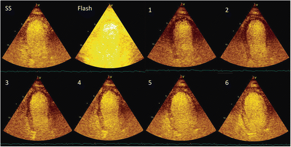
Fig. 1
Fig. 1
Normal myocardial contrast perfusion echocardiograph: qualitative assessment. Ultrasound contrast infused until steady state achieved (SS – steady state). A ‘flash’ of high mechanical index ultrasound destroys the contrast microbubbles within the imaging beam. Assessment of myocardial perfusion is then made as the microbubbles return to the myocardium over subsequent cardiac cycles (1–6). Normal replenishment occurs over 5–6 cardiac cycles at rest, 2–3 cardiac cycles with stress
Fig. 2
Fig. 2
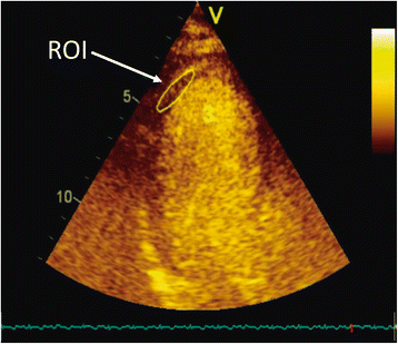
Qualitative assessment of myocardial perfusion involves specification of a region of interest (ROI) classically corresponding to individual left ventricular myocardial segments
Fig. 3
Fig. 3
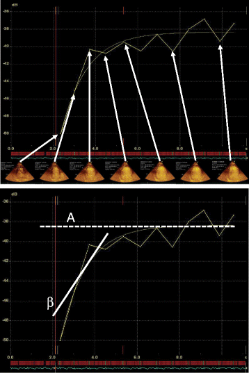
Myocardial contrast perfusion echocardiography (MCPE) quantitative assessment. Regions of interest (ROI) are defined and rate of change in signal intensity assessed at end‐diastolic frames. The plateau signal intensity (A) is considered to represent the myocardial capillary blood volume. The rate of replenishment (β) of the microbubbles is considered as the velocity of blood. The product of A  ×  β is considered to represent the myocardial blood flow
Fig. 4
Fig. 4
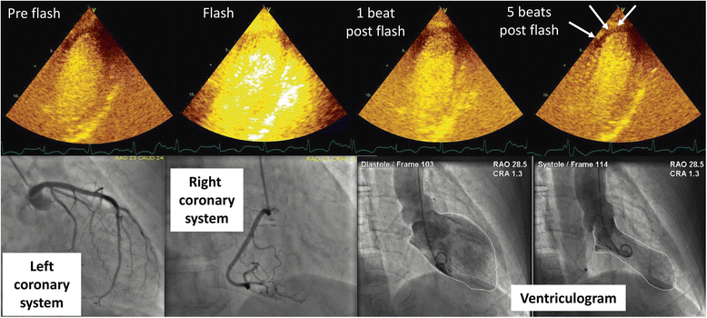
Takotsubo’s cardiomyopathy with microvascular dysfunction (arrows). Endocardial perfusion defect shown at 5 beats post flash in the apical region where transient apical hypokinesis was visualized. Coronary angiography confirmed normal vasculature and left ventriculography demonstrated apical ballooning

References

    1. Kurt M, Shaikh KA, Peterson L, et al. Impact of contrast echocardiography on evaluation of ventricular function and clinical management in a large prospective cohort. J Am Coll Cardiol. 2009;53:802–810. doi: 10.1016/j.jacc.2009.01.005. - DOI - PubMed
    1. Gramiak R, Shah PM, Kramer DH. Ultrasound cardiography: contrast studies in anatomy and function. Radiology. 1969;92:939–948. doi: 10.1148/92.5.939. - DOI - PubMed
    1. Keller MW, Feinstein SB, Watson DD. Successful left ventricular opacification following peripheral venous injection of sonicated contrast agent: an experimental evaluation. Am Heart J. 1987;114:570–575. doi: 10.1016/0002-8703(87)90754-X. - DOI - PubMed
    1. Feinstein SB, Cheirif J, Ten Cate FJ, et al. Safety and efficacy of a new transpulmonary ultrasound contrast agent: initial multicenter clinical results. J Am Coll Cardiol. 1990;16:316–324. doi: 10.1016/0735-1097(90)90580-I. - DOI - PubMed
    1. Lindner JR, Dent JM, Moos SP, et al. Enhancement of left ventricular cavity opacification by harmonic imaging after venous injection of Albunex. Am J Cardiol. 1997;79:1657–1662. doi: 10.1016/S0002-9149(97)00217-8. - DOI - PubMed

LinkOut - more resources