. 2016 Mar 29;113(13):E1806-15.
doi: 10.1073/pnas.1523575113. Epub 2016 Mar 14.

Aurone synthase is a catechol oxidase with hydroxylase activity and provides insights into the mechanism of plant polyphenol oxidases

Affiliations

Aurone synthase is a catechol oxidase with hydroxylase activity and provides insights into the mechanism of plant polyphenol oxidases

Christian Molitor et al. Proc Natl Acad Sci U S A. .

Abstract

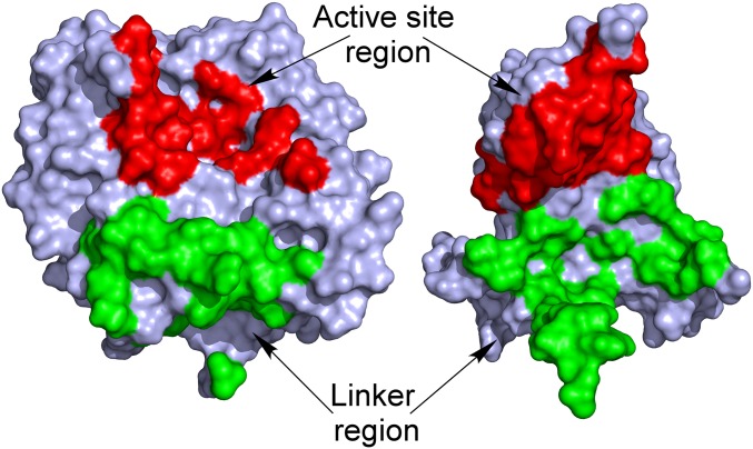
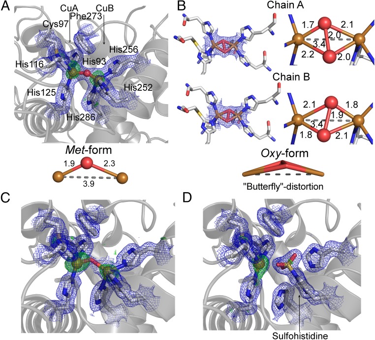
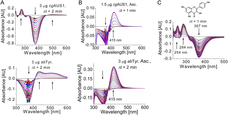
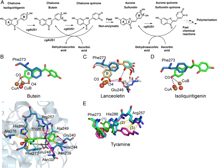
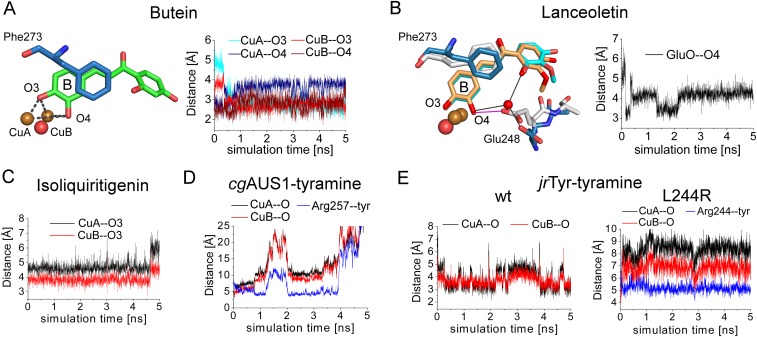
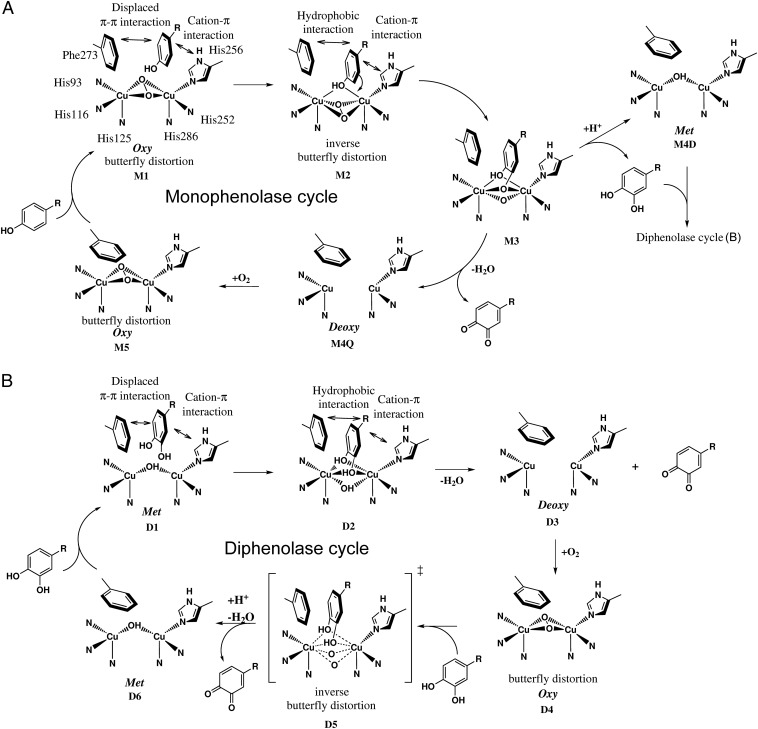
Tyrosinases and catechol oxidases belong to the family of polyphenol oxidases (PPOs). Tyrosinases catalyze theo-hydroxylation and oxidation of phenolic compounds, whereas catechol oxidases were so far defined to lack the hydroxylation activity and catalyze solely the oxidation of o-diphenolic compounds. Aurone synthase from Coreopsis grandiflora (AUS1) is a specialized plant PPO involved in the anabolic pathway of aurones. We present, to our knowledge, the first crystal structures of a latent plant PPO, its mature active and inactive form, caused by a sulfation of a copper binding histidine. Analysis of the latent proenzyme's interface between the shielding C-terminal domain and the main core provides insights into its activation mechanisms. As AUS1 did not accept common tyrosinase substrates (tyrosine and tyramine), the enzyme is classified as a catechol oxidase. However, AUS1 showed hydroxylase activity toward its natural substrate (isoliquiritigenin), revealing that the hydroxylase activity is not correlated with the acceptance of common tyrosinase substrates. Therefore, we propose that the hydroxylase reaction is a general functionality of PPOs. Molecular dynamics simulations of docked substrate-enzyme complexes were performed, and a key residue was identified that influences the plant PPO's acceptance or rejection of tyramine. Based on the evidenced hydroxylase activity and the interactions of specific residues with the substrates during the molecular dynamics simulations, a novel catalytic reaction mechanism for plant PPOs is proposed. The presented results strongly suggest that the physiological role of plant catechol oxidases were previously underestimated, as they might hydroxylate their--so far unknown--natural substrates in vivo.

Keywords: catechol oxidase; crystal structure; mechanism; type III copper protein; tyrosinase.


Conflict of interest statement

The authors declare no conflict of interest.

Figures

Fig. 1.
Fig. 1.
Overall structures of latent and mature aurone synthase. (A) Top view of mature aurone synthase. The coloring was performed according to the secondary structure features (α-helices, green; 310-helices, yellow; β-sheets, blue), and the characteristic features of AUS1 are colored red (loop carrying the insertion V237ANG240) and magenta (residual C-terminal peptide). (B) Side view of the dimeric biological assembly of mature AUS1. The interacting residues are colored orange (monomer chain B) and magenta (monomer chain D). (C) Overall structure of latent aurone synthase. The features of the C-terminal domain are as follows: the three proteolytic cleavage sites, colored magenta and labeled as (1), (2), and (3); the linker region (orange) that connects the catalytically active domain (gray) and the shielding C-terminal domain (green); and the residual peptide of the C-terminal domain (blue) and the active site shielding Ile456 (plug residue) (red). (D) The active site is shielded by the C-terminal residue Ile456, which is responsible for the latency of the proenzyme and functions like a plug [2Fo-Fc electron density map (blue mesh) contoured at 1.0 σ].
Fig. S1.
Fig. S1.
Primary structure, secondary structure, and features of latent and mature AUS1.
Fig. S2.
Fig. S2.
Secondary structure features [assigned using DSSP (64); compare with Fig. S1] and superimposition of mature and latent AUS1. (A) Secondary structure features of mature AUS1 (α-helices, red tubes; 310-helices, yellow tubes; β-strands, blue). (B) Superposition of active (colored beige) and latent (catalytically active domain, blue; shielding C-terminal domain, cyan) AUS1 (rmsd of 0.47 Å between 338 atom pairs). The location of the loop carrying the insertion is different in the latent proenzyme (latent, yellow; active, red).
Fig. S3.
Fig. S3.
Size exclusion chromatogram of mature AUS1 [41.6 kDa (15)] on Superdex 200 Increase. A buffer containing 50 mM sodium citrate and 1.5 M sodium chloride, pH 5.4, was used, and a flow rate of 0.5 mL/min was applied. Enzymatic assays for active AUS1 were performed by monitoring the oxidation of 50 µM fisetin in 125 mM sodium citrate pH 5.5 at 280 nm in a total volume of 1 mL (15). (A) Full (Left) and magnified (Right) chromatogram of mature AUS1. The dimeric AUS1 eluted at 14.10 mL (estimated mass, 81.0 kDa), and the monomeric AUS1 eluted at 15.80 mL (estimated mass, 44.1 kDa). (B) The calibration was performed using thyroglobulin (669 kDa), apoferritin (443 kDa), beta-amylase (200 kDa), alkoholdehydrogenase (150 kDa), albumin (66 kDa), carbonic anhydrase (29 kDa), and lysozyme (14 kDa).
Fig. 2.
Fig. 2.
Interactions between the shielding C-terminal domain and the catalytically active domain of latent AUS1. The latent structure is segmented by its domains (left, catalytically active domain; right, shielding C-terminal domain), and their solvent-accessible surface is presented. The interdomain interactions are located at two distinct areas: in the vicinity of the active site (colored red) and in the vicinity of the linker region (colored green).
Fig. 3.
Fig. 3.
Copper binding site of AUS1. The 2Fo-Fc electron density map (blue mesh) is contoured at 1.0 σ, and the anomalous Fourier difference map in A, C, and D (green mesh) is contoured at 3.0 σ. (A) Native met-copper center of recombinantly expressed AUS1. (B) Oxy-copper center of recombinantly expressed AUS1 (μ-η22-peroxo geometry; crystal soaked in H2O2). The oxygen atoms are slightly unsymmetrically bound to the copper atoms, and the μ-η22 peroxo complex displays a butterfly distortion. (C) Copper binding site of active AUS1 (occupancy CuB, ∼0.55). For clarity, the sulfohistidine (occupancy, ∼0.45) is not shown. (D) Inactive AUS1 (occupancy sulfohistidine, 0.9).
Fig. S4.
Fig. S4.
HPLC–ESI–MS data of the peptide carrying the sulfohistidine. (A) MS1 data of the modified peptide possessing a neutral loss of 80 Da in MS2 spectra. The mass precision of the performed measurements is better than 3 ppm. The MS1 masses match to a sulfonation of the modified peptide, whereas a putative phosphorylation can be excluded. (B) Exemplary MS2 spectrum of 595.93 m/z (MS1) displaying a neutral loss of 80 Da. (C) MS3 spectrum of 569.6 m/z (MS2) originating from 595.93 m/z (MS1). (D) List of matched peptide fragments.
Fig. S4.
Fig. S4.
HPLC–ESI–MS data of the peptide carrying the sulfohistidine. (A) MS1 data of the modified peptide possessing a neutral loss of 80 Da in MS2 spectra. The mass precision of the performed measurements is better than 3 ppm. The MS1 masses match to a sulfonation of the modified peptide, whereas a putative phosphorylation can be excluded. (B) Exemplary MS2 spectrum of 595.93 m/z (MS1) displaying a neutral loss of 80 Da. (C) MS3 spectrum of 569.6 m/z (MS2) originating from 595.93 m/z (MS1). (D) List of matched peptide fragments.
Fig. S5.
Fig. S5.
Monophenolase activity of AUS1 and its comparison with mushroom tyrosinase (abTYR). The reaction medium (1 mL) contained 50 µM substrate (A and B, isoliquiritigenin; C, kaempferol) in 100 mM sodium phosphate pH 6.5. (A) Comparison of difference spectra of the reaction of isoliquiritigenin catalyzed by AUS1 (Top) and abTYR (Bottom) without a reducing agent. In contrast to the reaction catalyzed by mushroom tyrosinase, only an extremely weak and very broad absorption band at a higher wavelength than expected appears, although the disappearance of substrate is visible. The reason for the occurring different reaction products remains unclear. However, the comparison of the enzymes in the presence of a reducing agent (compare with B) indicates that the highly reactive quinoid intermediates and products are responsible for the observed differences. (B) Difference spectra of the hydroxylation reaction (indicated by an increasing and narrow absorption band at 415 nm) (15, 16) catalyzed by AUS1 (Top) and abTYR (Bottom) in the presence of 1 mM sodium ascorbate as the reducing agent to trap the highly reactive quinoid intermediates and products. (C) Difference spectra of the hydroxylation reaction of kaempferol catalyzed by AUS1 in the presence of 5 mM H2O2 (41).
Fig. 4.
Fig. 4.
Reactivity and in silico-obtained substrate–enzyme complexes of AUS1. (A) Reaction scheme of the hydroxylation of isoliquiritigenin in the presence of ascorbic acid to trap highly reactive quinoid intermediates and products. (B) MD simulation snapshot of AUS1 in complex with butein. The reactive oxygen atoms at the B-ring of butein (O3, O4) are almost symmetrically located between CuA and CuB. The enzyme–substrate interactions are visualized by magenta lines (hydrogen bonding), yellow dashes (hydrophobic interactions), cyan dashed lines (π–π stacking), red dashes (cation–π interactions), and gray lines (water bridges). (C) Superimposition of snapshots obtained from different MD simulations of AUS1 with lanceoletin as the substrate. Residue Glu248 coordinates an oxygen atom of the A-ring through the formation of a water bridge (blue sticks; water bridges, gray lines) or one reactive oxygen atom (O4, B-ring) directly (white sticks; hydrogen bonding, magenta lines). (D) MD simulation snapshot of AUS1 in complex with isoliquiritigenin. (E) Three snapshots [0 ps, green (1); 44 ps, cyan (2); and 344 ps, magenta (3)] of the MD simulation of AUS1 with tyramine. Hydrophobic interactions of tyramine with Arg257 are visualized by yellow dashes.
Fig. S6.
Fig. S6.
Distance plots of the performed MD simulations. (A) Distances of the reactive oxygen atoms of butein (O3 and O4) to both copper atoms of AUS1. (B) Distance of the carboxylic oxygen atom to the substrate’s copper-coordinating oxygen atom. (C) Distances of the reactive oxygen atom of isoliquiritigenin to the CuA and CuB atoms of AUS1. (D) Distances of the reactive oxygen atom of tyramine to the CuA and CuB atoms of AUS1. The centers of masses were used to determine the distance of tyramine to the side chain of Arg257 (blue line). (E) Distances of the reactive oxygen atom of tyramine to the CuA and CuB atoms of the wild type (Left) and the L244R mutant (Right) of jrTYR. The centers of masses were used to determine the distance of tyramine to the side chain of Arg244 (blue line).
Fig. 5.
Fig. 5.
Proposed catalytic reaction mechanism of plant PPOs. (A) Monophenolase cycle: The monophenolic substrate is guided to the binuclear copper site [oxy-form, Cu(II)2O2, μ-η22 peroxide complex] by displaced π–π interactions with the tilt out gate residue Phe273 and cation–π interaction with the CuB binding histidine His256 (M1). When the substrate binds to the copper atoms, hydrophobic interactions with the substrate’s para-substituent become important additionally. The reactive substrate oxygen atom binds equally to both copper ions (M2), and the peroxide ligand is transferred to an inverse butterfly distortion (ligand field molecular dynamical simulations, the Cu–O–O–Cu torsion angles of the peroxo ligand fluctuate by ±20°) (32). Nucleophilic attack of the Cu2O2 moiety by the substrate results either in an o-diphenolic product and the met-form [M4D, Cu(II)2OH] or in an o-quinone and the deoxy-form [M4Q, Cu(I)2]. During product release, the gate residue swings back to its preferred position. Finally, oxygen uptake closes the catalytic cycle (M5). (B) Diphenolase cycle: The principles of the diphenolase cycle are similar to the monophenolase cycle described in A. The reactive substrate oxygen atoms bind equally to both copper ions of the met-form (D1, D2), and the o-quinone is released after electron transfer to the binuclear copper site resulting in the deoxy-form (D3). Oxygen uptake results in the oxy-form (D4) and substrate binding results in an inverse butterfly distortion in the transition state (D5). The catalytic cycle is closed by the release of an o-quinone resulting in the met-form of the binuclear copper site.

References

    1. Mayer AM. Polyphenol oxidases in plants and fungi: Going places? A review. Phytochemistry. 2006;67(21):2318–2331. - PubMed
    1. Tran LT, Taylor JS, Constabel CP. The polyphenol oxidase gene family in land plants: Lineage-specific duplication and expansion. BMC Genomics. 2012;13:395. - PMC - PubMed
    1. Dirks-Hofmeister ME, Singh R, Leufken CM, Inlow JK, Moerschbacher BM. Structural diversity in the dandelion (Taraxacum officinale) polyphenol oxidase family results in different responses to model substrates. PLoS One. 2014;9(6):e99759. - PMC - PubMed
    1. King RS, Flurkey WH. Effects of limited proteolysis on broad bean polyphenoloxidase. J Sci Food Agric. 1987;41(3):231–240.
    1. Marusek CM, Trobaugh NM, Flurkey WH, Inlow JK. Comparative analysis of polyphenol oxidase from plant and fungal species. J Inorg Biochem. 2006;100(1):108–123. - PubMed

Publication types